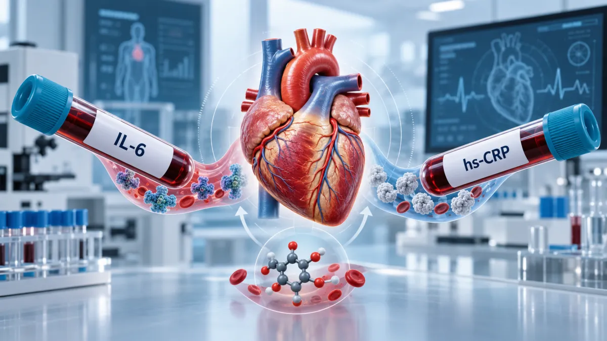
Can hs-CRP Miss Heart-Failure Risk in Type 2 Diabetes?
Introduction
Inflammation is an important link between type 2 diabetes and cardiovascular disease, but it cannot be reduced to a single laboratory marker. High-sensitivity C-reactive protein, or hs-CRP, is commonly used to estimate systemic inflammatory risk because it is accessible, relatively stable and supported by extensive cardiovascular research. A normal hs-CRP result, however, does not prove that every inflammatory pathway affecting the heart is inactive.
A new analysis published in JACC: Heart Failure raises that possibility. Investigators examined inflammatory biomarkers collected from participants in Look AHEAD, a large randomized study of adults with type 2 diabetes and overweight or obesity. Baseline hs-CRP was available for 1,902 participants, while interleukin-6, or IL-6, was measured in 1,507. Participants were then followed for approximately 12 years to determine who developed heart failure or atherosclerotic cardiovascular disease.
After accounting for traditional cardiovascular risk factors and statin use, elevated IL-6 remained associated with incident heart failure. hs-CRP, by contrast, was not independently associated with either heart failure or atherosclerotic cardiovascular events in this analysis. Among participants who did not already have cardiovascular disease, higher IL-6 was associated with nearly twice the subsequent risk of heart failure.
The distinction is biologically important. CRP is produced mainly by the liver in response to inflammatory signals, including IL-6. It therefore acts largely as a downstream indicator of inflammation. IL-6 participates more directly in immune signaling and can influence endothelial function, insulin resistance, vascular inflammation and tissue remodeling. Measuring CRP and measuring IL-6 are related, but they are not necessarily interchangeable views of the same process.
The study also found that the intensive lifestyle intervention used in Look AHEAD reduced IL-6 by 28.7% after one year, compared with an 11.8% reduction in participants receiving diabetes education and support. This does not prove that lowering IL-6 prevented heart failure, but it demonstrates that the biomarker responded substantially to changes involving nutrition, physical activity and weight management.
The findings do not establish IL-6 as a routine cardiovascular screening test, nor do they provide a validated concentration at which treatment should begin. The Look AHEAD ancillary study was observational within a randomized cohort: it identified an association, but it did not test whether ordering IL-6 or treating patients according to the result improves outcomes.
In this article, we will examine why heart failure is particularly relevant in type 2 diabetes, how IL-6 differs from hs-CRP, what the new study actually found and whether broader inflammatory testing can add useful information to conventional cardiometabolic risk assessment.
🎧 Listen to the Episode: IL-6 and the Hidden Risk of Heart Failure
Inflammation isn't one biological signal, and measuring one downstream marker can't necessarily capture everything happening across adipose tissue, blood vessels, kidneys, and the heart.
In this episode of The Health Pulse, we compare IL-6 and hsCRP through the lens of type 2 diabetes and heart failure risk. We explore evidence from the Look AHEAD population, explain why chronically elevated IL-6 is different from the temporary IL-6 surge produced during exercise, and examine why understanding cardiovascular risk requires looking beyond any single inflammation marker.
▶️ Click play below to listen, or keep reading to discover how insulin resistance, inflammation, kidney function, and cardiac stress can converge long before heart failure becomes obvious.
Why Diabetes Raises Heart-Failure Risk
Heart failure is sometimes assumed to be the final result of a heart attack or severely blocked coronary arteries. Those conditions can certainly damage the heart, but type 2 diabetes can increase heart-failure risk through several pathways even when a person has never experienced a recognized cardiovascular event.
Insulin resistance affects more than glucose control. It is commonly accompanied by elevated blood pressure, abdominal obesity, fatty liver, abnormal lipoproteins and chronic kidney disease. Together, these conditions increase the workload placed on the heart and promote fluid retention, vascular dysfunction and structural changes in the cardiac muscle.
The heart’s metabolism may also change. A healthy heart can move flexibly among fatty acids, glucose, lactate and ketones according to energy demands. Insulin resistance can reduce glucose uptake while increasing exposure to circulating fatty acids. When the supply of fatty acids exceeds the heart’s capacity to use or safely store them, lipid intermediates, oxidative stress and mitochondrial dysfunction may interfere with normal cardiac energy production.
Small blood vessels within the heart are another important part of the process. Endothelial dysfunction can reduce nitric oxide availability and impair the ability of the coronary microcirculation to respond to changing oxygen demands. This does not necessarily produce the focal obstruction seen in a major coronary artery, but it may contribute to chronic myocardial stress and impaired relaxation.
Inflammation connects many of these abnormalities. Cytokines such as IL-6 can participate in endothelial activation, immune-cell recruitment and signaling that promotes cardiac fibrosis. As fibrous tissue accumulates, the ventricle may become stiffer and less able to relax and fill normally. This is particularly relevant to heart failure with preserved ejection fraction, or HFpEF, which is strongly associated with obesity, hypertension, insulin resistance and type 2 diabetes.
Kidney dysfunction can amplify the problem. Reduced filtration, urinary albumin loss and impaired sodium handling can increase fluid volume and blood pressure. The heart and kidneys then place additional stress on one another, creating a cardiorenal cycle that can progress before obvious symptoms appear.
These pathways help explain why an assessment based only on LDL cholesterol or obstructive coronary disease may underestimate heart-failure risk in someone with diabetes. Glucose, insulin resistance, kidney health, blood pressure, inflammation and body composition all contribute to the environment in which heart failure develops.
The new Look AHEAD analysis is important within this context because IL-6 may capture part of the inflammatory and metabolic burden connected more closely with heart failure than with atherosclerotic events. The finding does not establish causation, but it supports the idea that heart-failure risk in type 2 diabetes is broader than plaque formation alone.
Key takeaway: Type 2 diabetes can promote heart failure through insulin resistance, altered cardiac metabolism, endothelial dysfunction, kidney disease and inflammation—even without a previous heart attack. This may be why a signaling cytokine such as IL-6 revealed risk that hs-CRP did not capture in the new analysis.
IL-6 and hs-CRP Measure Different Parts of Inflammation
IL-6 and hs-CRP are connected, but they are not the same kind of biomarker. IL-6 is a cytokine—a signaling protein released by different tissues to coordinate immune, metabolic and repair responses. CRP is an acute-phase protein produced primarily by the liver after it receives inflammatory signals, particularly from IL-6.
This places the two markers at different points in the inflammatory cascade. IL-6 is considered relatively upstream because it participates in generating and directing the response. CRP sits farther downstream and reflects how strongly the liver has responded to the combined inflammatory signals reaching it. The relationship is sometimes summarized as a pathway moving from IL-1 to IL-6 and then to hepatic CRP production.
IL-6 can be released by immune cells, vascular tissue, adipose tissue and other organs. In obesity and insulin resistance, dysfunctional adipose tissue can attract macrophages and produce inflammatory signals that extend beyond the fat depot. Chronic elevation may contribute to hepatic insulin resistance, endothelial activation and changes in the structure and function of the heart.
However, IL-6 should not be described as universally harmful. Its effects depend on its source, duration, concentration and signaling pathway. Contracting skeletal muscle can release IL-6 temporarily during exercise, where it participates in energy regulation and may be followed by anti-inflammatory effects. A short exercise-related rise is biologically different from persistent IL-6 signaling associated with visceral obesity, infection or chronic disease.
High-sensitivity CRP measures very small concentrations of CRP and is better standardized for routine clinical use. It can help identify residual inflammatory cardiovascular risk, particularly when interpreted with cholesterol, ApoB, blood pressure, smoking status and other established risk factors. It remains useful even though it cannot identify the source or exact pathway producing the inflammation.
CRP is also nonspecific. An infection, injury, autoimmune flare, recent surgery or intense exercise can temporarily increase it. Because it represents a downstream liver response, a lower result does not necessarily mean that every upstream cytokine or tissue-specific inflammatory process is quiet. The new Look AHEAD findings suggest that this distinction may be relevant to heart failure.
IL-6 testing has limitations of its own. Concentrations are lower, more biologically variable and less standardized than hs-CRP. An elevated result can occur with infection, autoimmune disease, cancer, obesity and many other conditions. There is no universally accepted IL-6 threshold for predicting heart failure, and a single measurement cannot identify why the cytokine is elevated.
The new JACC: Heart Failure analysis does not show that hs-CRP has become obsolete. It shows that the two markers may carry different information. In this population with type 2 diabetes and overweight or obesity, baseline IL-6 retained an association with future heart failure after adjustment, while hs-CRP did not.
Key takeaway: IL-6 is an upstream signaling cytokine, while hs-CRP is primarily a downstream hepatic response. They overlap, but they do not provide identical information—and neither marker should be interpreted as a complete measurement of inflammation by itself.
What the Study Found
The new research was an ancillary analysis of Look AHEAD, a large multicenter randomized trial originally designed to test whether an intensive lifestyle intervention could improve cardiovascular outcomes in adults with type 2 diabetes and overweight or obesity. The inflammatory analysis used stored baseline blood samples and long-term clinical follow-up from a subset of participants.
Researchers had baseline hs-CRP measurements for 1,902 participants and IL-6 measurements for 1,507. The group with hs-CRP results had an average age of approximately 58 years, and nearly 58% were women. Median baseline concentrations were 4.04 mg/L for hs-CRP and 2.07 pg/mL for IL-6.
The investigators examined whether either biomarker was associated with subsequent atherosclerotic cardiovascular disease or incident heart failure. Their statistical models accounted for demographic characteristics, conventional cardiovascular risk factors and statin use. They also evaluated participants separately according to whether cardiovascular disease was already present at baseline.
After these adjustments, hs-CRP was not significantly associated with either incident heart failure or atherosclerotic cardiovascular disease. This does not mean hs-CRP has no cardiovascular value in other populations. It means that within this particular cohort and statistical analysis, baseline hs-CRP did not provide an independent association with the outcomes after the other measured risk factors were considered.
IL-6 produced a different result. Across the overall cohort, each one-standard-deviation increase in log-transformed IL-6 was associated with a 43% higher risk of developing heart failure. The confidence interval ranged from 16% to 75%, supporting a statistically significant association.
The relationship was stronger among participants without known cardiovascular disease. In that group, each one-standard-deviation increase in log IL-6 was associated with a 92% higher risk of incident heart failure. This is the basis for the statement that higher IL-6 was linked to nearly twice the risk—not a finding that every person above one specific IL-6 concentration had exactly double the risk.
The analysis did not find the same independent relationship between IL-6 and atherosclerotic cardiovascular disease. That distinction is notable because it suggests IL-6 may have been capturing inflammatory processes more closely connected with the development of heart failure than with conventional plaque-related events in this population.
The randomized lifestyle component provided a second finding. After one year, participants assigned to intensive lifestyle intervention experienced a 28.7% reduction in IL-6, compared with an 11.8% reduction among those receiving diabetes education and support. The difference was statistically significant.
This result shows that IL-6 was modifiable, but it does not demonstrate that the reduction itself prevented heart failure. The intervention changed weight, nutrition, physical activity and other metabolic factors simultaneously. The study therefore cannot determine whether lowering IL-6 caused a clinical benefit or simply reflected broader metabolic improvement.
The published analysis compared the biomarkers’ associations with outcomes; it was not a clinical trial assigning patients to IL-6-guided versus hs-CRP-guided care. Describing IL-6 as “better” should therefore be limited to its stronger independent association with incident heart failure in this dataset—not interpreted as proof that it is already the superior screening test.
Key takeaway: IL-6 remained independently associated with future heart failure, particularly in participants without established cardiovascular disease, while hs-CRP did not. The finding is clinically intriguing but does not yet establish an IL-6 screening threshold or prove that lowering IL-6 prevents heart failure.
Why Could hs-CRP Miss the Signal?
The study does not establish exactly why IL-6 was associated with heart failure while hs-CRP was not. Several biological and statistical explanations are possible, but they should be treated as hypotheses rather than conclusions proven by the analysis.
One possibility is that hs-CRP was already elevated across much of the study population. The median baseline concentration was 4.04 mg/L, consistent with a substantial background inflammatory burden. Because all participants had type 2 diabetes and overweight or obesity, hs-CRP may have reflected shared metabolic inflammation without distinguishing as clearly between those who would and would not later develop heart failure.
IL-6 sits closer to the signaling processes that generate the acute-phase response. It can be produced by dysfunctional adipose tissue, activated immune cells and vascular tissue, and it may participate in endothelial dysfunction, myocardial fibrosis and adverse cardiac remodeling. CRP integrates these and other signals through the liver but does not identify which tissue or inflammatory pathway initiated them.
This difference could make IL-6 more sensitive to inflammation connected specifically with the development of heart failure. A downstream marker such as CRP may still indicate general cardiovascular risk while providing less information about the particular cytokine activity influencing the myocardium and microvasculature.
Liver biology may also affect the relationship. CRP production depends on the liver’s response to IL-6 and other signals. Type 2 diabetes commonly coexists with metabolic liver disease, which could theoretically influence how upstream inflammation is translated into circulating CRP. The Look AHEAD analysis did not establish liver dysfunction as the reason for the discordance, but it illustrates why one downstream protein cannot represent every component of systemic inflammation.
Medications add another layer. Statins can reduce hs-CRP independently of their effect on LDL cholesterol. The researchers adjusted their models for statin use, but statistical adjustment cannot capture every difference in medication intensity, adherence or duration. Other diabetes, blood-pressure and anti-inflammatory treatments may also influence biomarkers or heart-failure risk.
The two biomarkers were not available in identical groups. hs-CRP was measured in 1,902 participants, while IL-6 was measured in 1,507. Differences in sample availability and participant characteristics can influence results. The study also relied on one baseline measurement, which cannot distinguish persistent inflammation from temporary changes caused by infection, injury, exercise or another short-lived condition.
Most importantly, the analysis was not designed as a diagnostic competition between IL-6 and hs-CRP. It did not establish clinical cutoffs, compare screening algorithms or demonstrate that adding IL-6 improved patient classification beyond established heart-failure assessments. Its conclusion is narrower: IL-6 retained an independent association with incident heart failure in this dataset, while hs-CRP did not.
The finding should therefore expand how inflammation is understood rather than eliminate hs-CRP. The 2025 ACC scientific statement on inflammation and cardiovascular disease continues to recognize hs-CRP as the best-established inflammatory biomarker for cardiovascular risk assessment. The new study suggests that heart failure may require a broader inflammatory lens than atherosclerotic disease alone.
Key takeaway: hs-CRP may have reflected broad metabolic inflammation without isolating the pathways most closely connected with heart failure. IL-6 showed a stronger association, but the study does not prove why the markers diverged or establish that IL-6 should replace hs-CRP.
Why Lifestyle Lowered IL-6
The intensive lifestyle intervention used in Look AHEAD was not a vague recommendation to “eat better and exercise.” It was a structured program designed to produce and maintain meaningful weight loss through calorie reduction, increased physical activity and frequent behavioral support.
Participants were initially encouraged to lose at least 7% of their body weight and gradually work toward approximately 175 minutes of moderate-intensity physical activity each week. The program included regular counseling, meal planning, self-monitoring and, during the early phase, optional meal-replacement products to make calorie targets easier to achieve.
At one year, the intensive group had lost an average of approximately 8.6% of its starting body weight, compared with less than 1% in the diabetes education and support group. In the new inflammatory analysis, this was accompanied by a 28.7% reduction in IL-6, compared with an 11.8% decline in the comparison group.
Loss of dysfunctional adipose tissue is one likely explanation. Fat tissue is not simply a passive calorie-storage compartment. As adipocytes enlarge and exceed their safe storage capacity, the tissue can become poorly oxygenated, attract immune cells and release more inflammatory mediators. Reducing fat mass may relieve some of this immune activation and decrease chronic cytokine production.
Improved insulin sensitivity may contribute as well. Lower insulin resistance can reduce excessive nutrient flow into the liver, improve glucose regulation and limit metabolic signals associated with oxidative stress. Changes in blood pressure, sleep, liver fat and physical fitness may also influence the inflammatory environment.
Exercise adds an important nuance. Working skeletal muscle can temporarily release IL-6 during physical activity. This short-lived myokine response helps coordinate energy availability and can stimulate anti-inflammatory signaling. It is biologically different from persistently elevated resting IL-6 associated with visceral obesity, immune activation and chronic metabolic disease. Regular physical activity can therefore produce a brief rise during exercise while contributing to lower basal inflammation over time.
The intervention changed several variables at once, making it impossible to determine how much of the IL-6 reduction came from weight loss, calorie restriction, food composition, exercise or improved metabolic health. The study also did not establish that lowering IL-6 was the mechanism responsible for any improvement in heart-failure risk.
That caution matters because the original Look AHEAD cardiovascular outcomes trial did not significantly reduce its primary composite cardiovascular endpoint, despite producing greater weight loss and improvements in several risk factors. A favorable biomarker change does not automatically translate into fewer clinical events.
The IL-6 result is still meaningful. It demonstrates that a structured lifestyle intervention can modify an inflammatory signal associated with future heart-failure risk. It also suggests that metabolic improvement may be visible through pathways that are not fully represented by glucose, weight or hs-CRP alone.
The practical objective should not be to chase the lowest possible IL-6 measurement. It should be to reduce the metabolic conditions capable of producing chronic inflammation: excess visceral fat, insulin resistance, poor glucose regulation, physical inactivity and declining muscle health.
Key takeaway: Intensive lifestyle intervention reduced IL-6 substantially more than diabetes education alone, probably because it changed weight, adipose-tissue function, insulin sensitivity and physical activity together. The reduction is encouraging, but the study does not prove that lowering IL-6 itself prevents heart failure.
Should IL-6 Be Tested Routinely?
The new findings are not enough to make IL-6 a routine cardiovascular screening test. An association with future heart failure is an important research signal, but a useful clinical test must do more than correlate with an outcome. It needs standardized measurement, validated decision thresholds and evidence that acting on the result improves patient care.
IL-6 currently lacks a universally accepted cardiovascular-risk range. Concentrations can differ among laboratory methods, and results may change with infection, autoimmune activity, injury, recent strenuous exercise or other temporary conditions. A value associated with greater risk across a research population cannot automatically determine an individual patient’s prognosis.
The Look AHEAD study also analyzed IL-6 as a continuous variable after logarithmic transformation. The reported 92% higher risk among participants without established cardiovascular disease applied to each one-standard-deviation increase in log IL-6. It was not based on a simple clinical boundary separating “safe” from “dangerous” results.
Clinical actionability remains another limitation. If IL-6 is elevated, there is no approved heart-failure prevention protocol based specifically on that concentration. The appropriate response would still be to investigate and manage established risks such as blood pressure, glucose regulation, kidney disease, obesity, physical inactivity and existing cardiovascular disease.
hs-CRP remains better established for assessing residual inflammatory risk related primarily to atherosclerotic cardiovascular disease. The 2025 American College of Cardiology scientific statement identifies hs-CRP as the preferred inflammatory analyte in selected clinical settings. It is standardized, widely available and supported by substantially more outcomes research than IL-6.
For heart-failure assessment, BNP and NT-proBNP are more clinically actionable than either hs-CRP or IL-6. These natriuretic peptides are released when the heart experiences increased wall stress. The 2026 ADA Standards of Care recommend considering BNP or NT-proBNP screening in adults with diabetes to help identify preclinical heart failure. An abnormal result may lead to echocardiography and more specific cardiovascular evaluation.
The biomarkers answer different questions. hs-CRP estimates part of the inflammatory risk associated with atherosclerosis. IL-6 may provide information about upstream inflammatory signaling. BNP and NT-proBNP reflect cardiac wall stress more directly. None of them independently diagnoses the cause or subtype of heart failure.
Traditional cardiometabolic testing remains essential. HbA1c and fasting glucose assess glycemic exposure, while fasting insulin can add context about insulin resistance. Creatinine, eGFR and urine albumin-to-creatinine ratio evaluate the kidney-heart connection. A lipid panel, ApoB, blood pressure and clinical history help determine atherosclerotic risk.
IL-6 may become useful for selected high-risk patients, specialty evaluation or enrollment in trials of inflammation-targeted therapies. Before routine use can be justified, prospective studies must show that adding IL-6 meaningfully improves risk classification and leads to an intervention that reduces heart-failure events.
Ordering more biomarkers is not automatically better testing. The most valuable measurement is one that answers a defined clinical question and leads to a clear next step.
Key takeaway: IL-6 is a promising research biomarker, but it is not yet a routine heart-failure screening test. hs-CRP remains better established for inflammatory cardiovascular risk, while BNP or NT-proBNP is more directly actionable when evaluating heart-failure risk in people with diabetes.
A More Complete Heart-Risk Assessment
The main lesson from the IL-6 study is not that every person with type 2 diabetes needs another inflammatory test. It is that cardiovascular risk cannot be represented by one biomarker. A useful assessment should examine the metabolic drivers, vascular risk, kidney function, inflammation and evidence of cardiac stress together.
HbA1c and fasting glucose establish the degree of glycemic exposure, but neither shows how much insulin the body requires to maintain that glucose. Fasting insulin can provide additional context about insulin resistance, particularly when glucose still appears acceptable. Persistently elevated insulin may indicate continued metabolic stress before diabetes becomes visibly worse.
A lipid panel helps identify elevated triglycerides, low HDL cholesterol and other patterns commonly associated with insulin resistance. ApoB estimates the number of atherogenic particles capable of entering the arterial wall. This remains important because the new IL-6 study focused on heart failure, while people with diabetes also face substantial risk from coronary artery disease and stroke.
Kidney testing deserves equal attention. Creatinine and estimated glomerular filtration rate assess filtration, while a urine albumin-to-creatinine ratio can reveal vascular and glomerular injury before kidney function declines substantially. Kidney disease increases heart-failure risk through fluid retention, blood-pressure changes, vascular dysfunction and shared inflammatory pathways.
hs-CRP can add an established measure of systemic inflammatory risk, but it should ideally be tested when the person is metabolically stable and free from an obvious acute infection or injury. A markedly elevated or unexpected result may need to be repeated rather than immediately interpreted as chronic cardiovascular inflammation.
IL-6 may add research or specialist-level information in selected situations, but no validated cardiovascular decision threshold currently exists. An elevated value should not be used by itself to diagnose heart disease, predict exactly who will develop heart failure or justify an inflammation-targeting treatment.
BNP and NT-proBNP answer a more heart-specific question. These peptides rise when cardiac walls experience increased stress. The 2026 ADA Standards of Care recommend considering natriuretic-peptide screening in adults with diabetes to facilitate earlier identification of preclinical heart failure. An elevated result may support further assessment with electrocardiography, echocardiography or a cardiology evaluation.
Natriuretic peptides also require clinical interpretation. Age, kidney dysfunction and atrial fibrillation can raise the results, while obesity may suppress them. A low result can make clinically significant heart failure less likely in many circumstances, but no laboratory value should override persistent symptoms.
Shortness of breath, declining exercise tolerance, unexplained fatigue, swelling of the legs, rapid fluid-related weight gain or difficulty breathing while lying flat require clinical evaluation. Laboratory testing can guide the investigation, but it cannot replace physical examination and cardiac imaging when heart failure is suspected.
Quick Lab Mobile provides convenient at-home blood collection in Miami for HbA1c, fasting glucose, fasting insulin, comprehensive metabolic panels, lipid testing, ApoB, hs-CRP and other clinician-requested cardiovascular and metabolic markers. When ordered appropriately, BNP or NT-proBNP and specialty inflammatory testing can be incorporated into a broader assessment rather than interpreted in isolation.
Key takeaway: Cardiovascular assessment in type 2 diabetes should combine metabolic, lipid, kidney, inflammatory and cardiac-stress markers. IL-6 may add new information, but established tests such as ApoB, urine albumin, eGFR and BNP or NT-proBNP currently provide clearer clinical pathways.
Conclusion
The new Look AHEAD analysis adds an important layer to how inflammation is understood in type 2 diabetes. hs-CRP remains a useful and well-established cardiovascular biomarker, but it does not measure every inflammatory pathway that may affect the heart.
In this cohort of adults with type 2 diabetes and overweight or obesity, IL-6 remained independently associated with incident heart failure after adjustment for conventional cardiovascular risks and statin use. The relationship was strongest among participants without previously diagnosed cardiovascular disease, where higher IL-6 was associated with nearly twice the subsequent risk.
hs-CRP did not show an independent association with heart failure or atherosclerotic cardiovascular disease in the same analysis. This does not invalidate hs-CRP or establish IL-6 as its clinical replacement. It suggests that upstream cytokine signaling and downstream hepatic inflammation can reveal different parts of cardiometabolic risk.
The lifestyle findings were equally relevant. Intensive intervention reduced IL-6 by 28.7% after one year, compared with 11.8% under diabetes education and support. The program changed weight, nutrition, physical activity and metabolic health simultaneously, so the study cannot determine which component produced the reduction or whether lowering IL-6 directly prevented heart failure.
IL-6 is not yet ready for universal cardiovascular screening. It lacks standardized decision thresholds, can change in response to many conditions and has not been shown to improve outcomes when used to guide treatment. For most patients, established assessments—including blood pressure, HbA1c, fasting glucose, kidney function, urine albumin, lipid testing, ApoB, hs-CRP and appropriately selected BNP or NT-proBNP—remain more actionable.
The broader message is that inflammation is not a single number. Heart-failure risk in type 2 diabetes develops through the combined effects of insulin resistance, dysfunctional adipose tissue, kidney disease, endothelial dysfunction, altered cardiac metabolism and immune signaling. A normal hs-CRP result cannot exclude every one of these pathways.
Quick Lab Mobile offers at-home blood collection in Miami for cardiometabolic, inflammatory, kidney and cardiovascular biomarkers requested by a healthcare professional. Testing should be selected according to the clinical question and interpreted as part of the complete cardiovascular picture.
👉 Need a specimen collection?. Book Now
Disclaimer:
The information provided in this blog, podcast, and associated content is for educational and informational purposes only and is not intended as a substitute for professional medical advice, diagnosis, or treatment. The content shared is based on reputable sources, medical literature, and expert insights, but it should not be used as a replacement for direct consultation with a licensed healthcare provider.
No Doctor-Patient Relationship: Engaging with this content does not create a doctor-patient relationship between you and QuickLabMobile or any contributors. Always consult with a qualified physician, specialist, or healthcare professional before making any medical decisions, changing your treatment plan, or starting/stopping any medications.
Not a Substitute for Medical Advice: While we strive to provide accurate and up-to-date information, medicine is constantly evolving. New research, treatments, and medical recommendations may emerge, and individual health conditions can vary. Do not rely solely on this content for health decisions. If you are experiencing symptoms, have concerns about your health, or require medical assistance, seek immediate care from a licensed medical professional.
Emergency Situations: If you are experiencing a medical emergency, such as difficulty breathing, chest pain, signs of a stroke, or any other life-threatening condition, call 911 (or your local emergency services) immediately. Do not delay seeking emergency care based on information provided here.
Liability Disclaimer: QuickLabMobile, its contributors, and any associated entities do not assume liability for any damages, harm, or adverse outcomes resulting from the use, interpretation, or misuse of the information provided in this content. You are responsible for your own healthcare decisions and should always verify information with a trusted medical professional.
External Links & References: This content may include links to external sources, medical studies, or third-party websites for further reading. These links are provided for convenience and informational purposes only. QuickLabMobile does not endorse, control, or take responsibility for the accuracy of external content. Always verify information with authoritative sources such as the CDC, NIH, WHO.
Final Note: Your health is unique, and what works for one person may not be suitable for another. Stay informed, ask questions, and always prioritize professional medical guidance