Cortisol Rhythms and Muscle Insulin Resistance
Introduction
Insulin resistance is often described as though it were a single metabolic defect affecting the entire body equally. In reality, insulin acts differently in skeletal muscle, adipose tissue and the liver. One tissue can become strongly insulin resistant while another continues responding to insulin, producing metabolic patterns that may look very different despite sharing the same diagnosis.
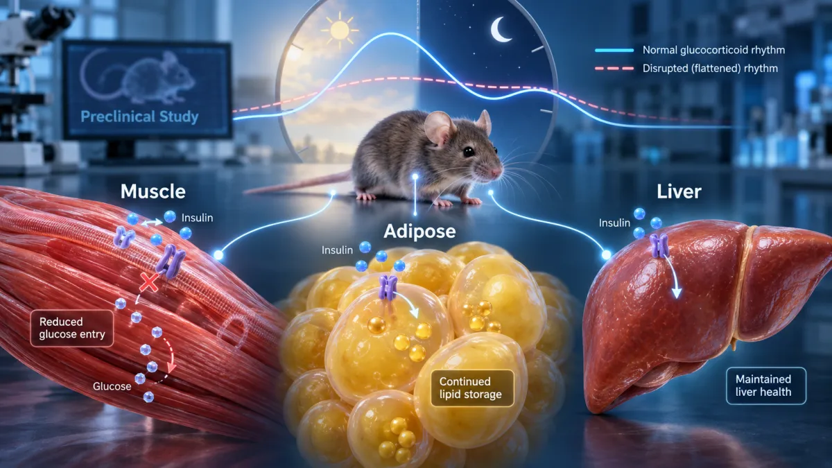
A new mouse study published in Cell Reports offers a striking example. Researchers disrupted the animals’ normal daily glucocorticoid rhythm without feeding them a high-fat diet. In mice, the primary glucocorticoid is corticosterone, which serves a role similar to cortisol in humans. Rather than simply increasing the hormone at one moment, the intervention flattened its normal rise-and-fall pattern across the day.
The animals gained fat and developed hyperinsulinemia and insulin resistance. However, this was not the same metabolic phenotype normally produced by diet-induced obesity. The most severe insulin resistance developed in skeletal muscle—the tissue responsible for clearing a substantial portion of glucose from the bloodstream after a meal.
Adipose tissue remained comparatively responsive to insulin and continued storing energy. Because fat tissue retained this capacity, circulating lipid was less likely to overflow into the liver. The animals consequently developed obesity without the pronounced hepatic steatosis commonly observed in mice fed a high-fat diet.
The findings challenge the idea that obesity, insulin resistance and fatty liver always progress together through one predictable pathway. They suggest that disrupted glucocorticoid timing can redistribute insulin resistance among different organs, producing a form of metabolic dysfunction dominated by impaired muscle glucose disposal rather than adipose failure and liver-fat accumulation.
This distinction could eventually help explain why people with similar body weights or glucose levels can have very different metabolic profiles. One person may have substantial fatty liver and dysfunctional adipose tissue, while another may have relatively little liver fat but severe muscular insulin resistance.
However, the study was conducted in mice. It does not establish that disrupted cortisol rhythms produce the same tissue-specific pattern in humans, nor does it make a single cortisol blood test a diagnostic test for insulin resistance. Instead, it provides a mechanistic framework for understanding why the timing of hormonal signals—and not merely their average concentration—may matter for metabolic health.
In this article, we will examine how normal cortisol rhythms work, what happened when researchers flattened them, why skeletal muscle and adipose tissue responded differently, and what these preliminary findings may eventually mean for evaluating insulin resistance in people.
🎧 Listen to the Episode: Cortisol Rhythms and Insulin Resistance
Insulin resistance isn't one uniform state affecting every organ simultaneously. Muscle, liver, and adipose tissue can respond differently—and emerging experimental research suggests that the timing of hormonal signals may help determine where metabolic dysfunction appears.
In this episode of The Health Pulse, we explore a fascinating preclinical study that flattened normal glucocorticoid rhythms without simply increasing total hormone exposure. The resulting pattern—substantial fat gain, pronounced skeletal-muscle insulin resistance, yet relative protection from fatty liver—challenges the idea that every form of insulin resistance follows the same metabolic pathway.
▶️ Click play below to listen, or keep reading to discover what this research can—and cannot yet—tell us about cortisol rhythms, circadian health, hyperinsulinemia, and metabolic disease in humans.
Why Cortisol Is Supposed to Rise and Fall
Cortisol is not meant to remain constant throughout the day. In humans, secretion normally rises during the early morning, increases further around awakening and then gradually declines toward its lowest concentration at night. Superimposed on this daily pattern are smaller pulses that allow tissues to experience alternating periods of glucocorticoid exposure and relative recovery.
This rhythm is coordinated by the hypothalamic-pituitary-adrenal axis and synchronized with the body’s central circadian clock. Light exposure, sleep, meals, physical activity and psychological stress can all influence its timing. Cortisol helps prepare the body for activity by increasing fuel availability, supporting blood pressure and influencing immune function.
Its metabolic effects are especially important. Cortisol can stimulate hepatic glucose production, reduce insulin-mediated glucose uptake in skeletal muscle and mobilize stored energy during fasting or stress. These actions are adaptive when they occur at the appropriate time. They help maintain glucose availability overnight and provide additional fuel when the body must respond to a physical or psychological demand.
Problems may develop when the rhythm becomes flattened. A flattened pattern does not necessarily mean cortisol is extremely elevated all day. It can instead mean that the normal morning peak is reduced, the nighttime concentration remains higher than expected, or the difference between the high and low points becomes smaller.
That distinction matters because tissues respond not only to the amount of glucocorticoid they receive but also to when the signal arrives. Glucocorticoid receptors interact with cellular clocks and influence the expression of genes involved in glucose transport, mitochondrial function, lipid metabolism and insulin signaling. Continuous exposure may therefore produce a different response than brief, properly timed peaks—even when total daily exposure is similar.
In the new mouse study, researchers implanted corticosterone pellets that flattened the animals’ normal hormone rhythm. Because mice are nocturnal, the timing of their natural corticosterone cycle differs from the human cortisol cycle, but the underlying principle is similar: glucocorticoid signaling normally changes predictably across the day.
Flattening that signal removed the separation between periods of higher and lower exposure. The result was not simply a miniature version of Cushing syndrome or ordinary diet-induced obesity. It produced a distinct redistribution of insulin resistance, with skeletal muscle affected much more severely than adipose tissue.
This also explains why a single morning cortisol measurement provides limited information about circadian regulation. One blood sample may detect marked cortisol excess or deficiency, but it cannot show whether the full daily rhythm is appropriately timed and sufficiently variable.
Key takeaway: Cortisol is a rhythmic metabolic signal, not merely a number that should be high or low. Losing its normal daily rise and fall may affect insulin signaling differently from a brief stress response or conventional diet-induced obesity.
Separating Hormone Timing From Diet
To determine whether glucocorticoid timing could produce its own metabolic phenotype, the researchers varied hormone rhythm and diet independently. The mice had either a normal or flattened corticosterone rhythm and consumed either standard laboratory food or a high-fat diet.
This created four experimental conditions:
Normal glucocorticoid rhythm with a standard diet
Flattened glucocorticoid rhythm with a standard diet
Normal glucocorticoid rhythm with a high-fat diet
Flattened glucocorticoid rhythm with a high-fat diet
The design allowed the investigators to distinguish changes caused by hormone timing from those caused by diet. It also showed what happened when both metabolic stressors were present simultaneously.
The corticosterone intervention raised hormone concentrations during the animals’ normal rest period while reducing the usual peak near the beginning of their active period. Importantly, this flattened the rhythm without substantially increasing average corticosterone exposure. The experiment was therefore testing the loss of hormonal timing more than conventional glucocorticoid excess.
After 30 days, mice eating the high-fat diet had approximately three times as much fat mass as the control animals. Mice with flattened glucocorticoid rhythms eating the standard diet accumulated nearly as much—approximately 2.5 times the control level. Animals exposed to both interventions gained the most fat, suggesting that diet and hormone-rhythm disruption acted through partly independent and additive mechanisms. Weill Cornell Medicine
The rhythm-disrupted mice initially lost some lean mass, but that loss occurred mainly during the first week. Lean mass then stabilized and began increasing. Their continued fat gain therefore could not be explained simply by progressive muscle wasting.
The most surprising difference appeared in the liver. High-fat-fed mice developed substantial hepatic fat accumulation, as expected. The glucocorticoid-flattened mice became similarly obese while storing most of the additional lipid in subcutaneous and visceral white adipose tissue. Their livers remained comparatively protected from steatosis.
This comparison demonstrates why body weight alone cannot describe metabolic health. Two animals can accumulate similar amounts of body fat while directing glucose and lipids into different tissues and developing very different patterns of insulin resistance.
Key takeaway: By separating diet from glucocorticoid timing, the study produced two forms of obesity with similar fat gain but different metabolic consequences. Flattened hormone rhythms drove obesity largely independently of a high-fat diet and without producing the same degree of fatty liver.
Insulin Resistance Was Concentrated in Muscle
Skeletal muscle is one of the body’s largest destinations for glucose after a meal. When insulin rises, it signals muscle cells to move GLUT4 glucose transporters toward the cell membrane, allowing circulating glucose to enter the tissue for immediate energy production or storage as glycogen.
Because skeletal muscle represents such a large portion of body mass, impaired muscle glucose uptake can substantially reduce whole-body insulin sensitivity. The pancreas may then release more insulin to maintain normal glucose, creating hyperinsulinemia before fasting glucose or HbA1c becomes clearly abnormal.
The researchers used hyperinsulinemic-euglycemic clamps and glucose tracers to examine where insulin was—and was not—working. During a clamp, insulin is administered while glucose is carefully adjusted to keep blood sugar stable. The amount of glucose needed and the tissues absorbing the tracer provide a detailed picture of insulin sensitivity.
Mice with flattened glucocorticoid rhythms showed markedly reduced insulin-stimulated glucose uptake in major skeletal muscles, including the quadriceps and gastrocnemius. Their muscles were therefore failing to respond normally even while circulating insulin concentrations were very high.
This helps explain how the animals maintained relatively normal blood glucose despite severe muscular insulin resistance. The pancreas compensated by producing much more insulin. Glucose control was preserved temporarily, but only through a stronger hormonal signal.
That pattern resembles the normoglycemic hyperinsulinemic phase that can occur early in human insulin resistance. A person may have fasting glucose and HbA1c within conventional reference ranges while requiring unusually high insulin concentrations to keep glucose there. Normal glucose does not necessarily mean normal glucose regulation.
Adipose tissue showed a more complicated response. Insulin-stimulated glucose uptake was reduced in some fat depots, but insulin retained an important ability to suppress lipolysis—the release of stored fatty acids. This means adipose tissue was not completely insulin sensitive, but neither was it resistant to every action of insulin.
This selective response matters. With insulin continuing to restrain fat release, adipose tissue remained capable of holding much of the excess energy. The muscle could reject glucose while fat cells continued storing lipid, producing severe whole-body insulin resistance without the same rise in circulating fatty acids or liver-fat accumulation seen with diet-induced obesity.
Key takeaway: The study did not find uniform insulin resistance across the body. Skeletal muscle became profoundly resistant to insulin, while adipose tissue preserved insulin’s ability to suppress fat release. High insulin compensated for poor muscle glucose uptake and helped keep blood glucose relatively normal.
Why the Liver Remained Protected
Fatty liver commonly develops when adipose tissue can no longer safely store incoming energy. As fat cells enlarge and become insulin resistant, insulin loses some of its ability to suppress lipolysis. More fatty acids are then released into circulation and delivered to organs that were not designed to store large amounts of lipid.
The liver is one of the principal recipients of this overflow. It receives fatty acids released from adipose tissue, manufactures additional fat through de novo lipogenesis and packages triglycerides into lipoproteins for export. When lipid delivery and production exceed the liver’s ability to oxidize or export fat, triglycerides accumulate and hepatic steatosis develops.
This sequence is common in diet-induced obesity. Adipose tissue becomes inflamed and resistant to insulin, fatty-acid release increases, and excess lipid begins accumulating in the liver, skeletal muscle and pancreas. The problem is therefore not simply how much body fat exists, but whether adipose tissue can continue storing it appropriately.
The glucocorticoid-flattened mice followed a different path. Their adipose tissue preserved insulin’s ability to restrain lipolysis. Although the animals gained substantial fat mass, circulating lipids remained comparatively controlled because stored fatty acids were not being released as aggressively.
High insulin concentrations likely reinforced this containment. The pancreas was producing more insulin to compensate for poor glucose uptake by skeletal muscle, while adipose tissue remained responsive enough for that insulin to suppress fat release. Energy was therefore directed toward storage in subcutaneous and visceral fat rather than being redistributed extensively to the liver.
The liver also retained relatively normal insulin-mediated suppression of endogenous glucose production. This indicates that hepatic insulin resistance was much less pronounced than the resistance occurring in muscle. The animals consequently maintained normal circulating glucose and relatively little liver fat despite becoming obese and hyperinsulinemic.
This phenomenon illustrates the concept of adipose expandability. As long as adipose tissue can create or enlarge fat cells, maintain blood flow and respond to insulin, it can protect other organs by providing a relatively safe location for triglyceride storage. Once that capacity is exceeded or becomes dysfunctional, lipids begin spilling into ectopic tissues.
It also explains why two people with a similar BMI can have very different metabolic risks. One may store most excess energy in functional subcutaneous adipose tissue, while another may accumulate comparatively little body fat but develop substantial liver, muscle or pancreatic fat.
The findings should not be interpreted to mean that obesity is harmless when liver fat is absent. The rhythm-disrupted mice still had severe muscle insulin resistance and marked hyperinsulinemia. Their metabolic dysfunction had been redistributed, not eliminated.
Key takeaway: Adipose tissue protected the liver by remaining responsive to insulin’s suppression of fat release. Lipid stayed largely contained within fat tissue, allowing severe muscle insulin resistance and hyperinsulinemia to coexist with relatively little hepatic steatosis.
Insulin Resistance Is Not an On-Off Switch
Insulin resistance is often summarized as cells “not responding to insulin.” That description is useful but incomplete. Insulin controls multiple processes in several organs, and those processes do not necessarily become resistant at the same time or to the same degree.
In skeletal muscle, insulin promotes glucose uptake and glycogen storage. Muscle insulin resistance primarily reduces the disposal of glucose after meals. The pancreas may compensate by releasing more insulin, allowing glucose to remain normal during the earlier stages of dysfunction.
In adipose tissue, insulin suppresses the breakdown and release of stored triglycerides. It also supports glucose uptake and fat storage. These actions can become separated. In the glucocorticoid-flattened mice, insulin-stimulated glucose uptake was impaired in some adipose depots, yet insulin still suppressed fatty-acid release. The tissue was resistant to one action of insulin while remaining responsive to another.
The liver provides another example of selective insulin resistance. Insulin normally suppresses hepatic glucose production while promoting glycogen synthesis and influencing lipid metabolism. In common metabolic disease, the liver may continue producing too much glucose while insulin still supports de novo lipogenesis. The result can be the seemingly contradictory combination of hyperglycemia and continued liver-fat production.
This partial hepatic insulin resistance helps explain why high insulin does not necessarily shut down every insulin-regulated pathway equally. Some signaling branches become impaired, while others remain active or may even be overstimulated by compensatory hyperinsulinemia.
The new mouse study produced a different distribution. Muscle glucose uptake was severely impaired, but the liver continued suppressing glucose production in response to insulin and adipose tissue continued suppressing lipolysis. Whole-body testing could identify insulin resistance, but it would not reveal this internal distribution without tissue-specific measurements.
The driver of the disorder may influence where resistance appears first. Excess nutrient exposure, adipose dysfunction, physical inactivity, glucocorticoid treatment, disrupted circadian signaling, muscle loss and genetic factors may not produce identical metabolic phenotypes.
This has practical implications for interpreting laboratory results. Elevated fasting insulin with normal glucose may indicate successful pancreatic compensation for poor peripheral glucose disposal. Elevated triglycerides, fatty liver and rising ALT may suggest that adipose and hepatic dysfunction are also developing. A person can have substantial muscle insulin resistance without displaying every feature of metabolic syndrome.
No routine blood test can precisely measure insulin sensitivity separately in muscle, liver and adipose tissue. However, evaluating fasting glucose, fasting insulin, HbA1c, triglycerides, HDL cholesterol, liver markers, body composition and post-meal glucose patterns can provide clues about the dominant metabolic abnormalities.
Key takeaway: Insulin resistance can be organ-specific and pathway-specific. Muscle may resist glucose uptake while adipose tissue continues suppressing fat release and the liver remains responsive to insulin’s control of glucose production. The diagnosis describes a family of metabolic defects, not one uniform condition.
Could This Happen in Humans?
The mouse findings are biologically plausible for humans, but they should not be interpreted as proof that a flattened cortisol rhythm produces the same tissue-specific insulin resistance in people. Human metabolism, behavior and hormonal regulation are considerably more complex than a controlled animal model.
Altered cortisol rhythms have been reported in association with chronic psychological stress, rotating or overnight shift work, irregular sleep, sleep deprivation, depression, obstructive sleep apnea and chronic illness. Exogenous glucocorticoid medications can also change normal hypothalamic-pituitary-adrenal signaling, although their effects depend on the medication, dose, timing and duration.
Human observational research has connected features of the diurnal cortisol curve with obesity, waist circumference, insulin resistance and abnormal glucose regulation. The Multi-Ethnic Study of Atherosclerosis found associations between certain cortisol-curve characteristics and glycemia or insulin resistance. The Whitehall II cohort also linked altered diurnal cortisol patterns with impaired glucose metabolism and future diabetes risk.
These studies establish associations, not direction of causality. Insulin resistance and obesity may disturb the hypothalamic-pituitary-adrenal axis, while disrupted cortisol signaling may also worsen metabolism. Both could be influenced by sleep, medications, psychological stress, socioeconomic conditions, physical activity and meal timing.
The experimental manipulation in mice was also unusually precise. Researchers changed the corticosterone rhythm while independently controlling diet. In real life, circadian disruption rarely occurs alone. A night-shift worker may sleep less, eat at biologically inappropriate times, move less during daylight and experience greater stress. Those exposures can influence insulin sensitivity independently of cortisol.
Humans and mice also have different activity cycles. Mice are nocturnal and use corticosterone as their principal glucocorticoid, whereas humans are generally active during the day and primarily produce cortisol. The direction of the daily cycle must therefore be interpreted according to the species rather than transferred hour for hour.
The study also does not show that everyone with stress, poor sleep or an abnormal cortisol curve will develop obesity or muscle-dominant insulin resistance. Nor does it establish that correcting cortisol timing would reverse the phenotype in humans.
Its real value is conceptual. It demonstrates that glucocorticoid timing can independently influence where insulin resistance develops and where energy is stored. It provides a possible mechanism worth investigating in people whose metabolic profile does not follow the usual pattern of fatty liver, elevated triglycerides and generalized insulin resistance.
Key takeaway: Human studies connect altered cortisol rhythms with obesity and abnormal glucose regulation, but they do not yet confirm the tissue-specific pattern seen in these mice. The new findings are a mechanistic hypothesis for human research—not a clinical diagnosis or treatment protocol.
Can Cortisol Testing Detect This Pattern?
No currently validated clinical test can determine that a person has the same muscle-dominant insulin resistance produced in these mice. Cortisol testing can answer specific endocrine questions, but it cannot yet identify this experimental metabolic phenotype.
A morning serum cortisol measurement provides a snapshot near the time cortisol is normally elevated. It is useful in selected evaluations of adrenal insufficiency and other endocrine disorders, but a normal morning value does not prove that the complete daily rhythm is normal. It provides no information about whether cortisol falls appropriately during the evening.
Late-night salivary cortisol examines the opposite end of the curve. Cortisol should normally be very low late at night, so an elevated result can indicate loss of the expected nighttime nadir. It is one of the tests recommended by the Endocrine Society when evaluating appropriately selected patients for Cushing syndrome.
A 24-hour urinary free cortisol test estimates overall unbound cortisol production across an entire day. It may detect excessive exposure, but it largely averages away timing. Two people could have similar daily cortisol production while one has a strong morning-to-evening rhythm and the other has a much flatter curve.
The dexamethasone suppression test evaluates whether synthetic glucocorticoid feedback appropriately suppresses cortisol production. It is useful when hypercortisolism is suspected, but it does not directly reconstruct the normal circadian pattern.
Multiple salivary samples collected from awakening through bedtime can estimate the cortisol-awakening response and diurnal slope. This approach is frequently used in research, but sampling time, sleep schedule, eating, smoking, oral contamination, medications and adherence can substantially influence results. There are also no validated thresholds showing that a particular curve diagnoses muscle-specific insulin resistance.
Commercial four-point salivary cortisol panels are sometimes promoted as tests for “adrenal fatigue.” That term is not a recognized endocrine diagnosis, and these panels should not be used by themselves to diagnose adrenal disease, explain nonspecific symptoms or justify hormone treatment.
Testing should instead be selected according to the clinical question. Progressive features such as unexplained muscle weakness, easy bruising, wide purple stretch marks, osteoporosis, difficult-to-control hypertension or unusual fat redistribution may justify evaluation for true cortisol excess. Suspected adrenal insufficiency requires a different testing strategy.
Even a clearly abnormal cortisol pattern would not show where insulin resistance is located. Tissue-specific insulin sensitivity in research is assessed with techniques such as hyperinsulinemic-euglycemic clamps, glucose tracers, imaging and tissue analysis—not routine cortisol testing.
Key takeaway: A single morning cortisol cannot evaluate the full daily rhythm, and no cortisol panel can currently diagnose the muscle-dominant insulin resistance seen in this study. Validated cortisol tests should be chosen for specific endocrine questions and interpreted within the complete clinical context.
What Metabolic Testing Can Reveal
Routine laboratory testing cannot determine exactly how much insulin resistance is present in skeletal muscle, adipose tissue or the liver. It can, however, reveal whether the body is compensating for impaired insulin sensitivity and whether metabolic dysfunction is beginning to affect other organs.
Fasting glucose and HbA1c are useful for identifying abnormal glycemia, but they may remain normal while the pancreas is producing additional insulin. In the glucocorticoid-flattened mice, high insulin helped compensate for poor muscle glucose uptake. A similar compensatory state in humans could be missed if only glucose is measured.
Fasting insulin adds important context. An elevated result alongside normal glucose suggests that maintaining that glucose concentration requires greater pancreatic effort. As discussed in Quick Lab Mobile’s guide to fasting insulin, glucose can look reassuring while hyperinsulinemia is already present.
Fasting glucose and insulin can also be combined to estimate HOMA-IR. This calculation is convenient, but it is influenced heavily by fasting hepatic glucose regulation and cannot localize insulin resistance to muscle. A normal or mildly elevated HOMA-IR does not completely exclude impaired post-meal glucose disposal.
An oral glucose-tolerance test that measures both glucose and insulin provides more information about compensation. Glucose may remain within an acceptable range only because insulin rises excessively or stays elevated longer than expected. These insulin measurements are not included in a standard glucose-tolerance test unless specifically ordered.
Continuous glucose monitoring can show how glucose responds to meals, exercise, sleep and daily stress. It may reveal delayed glucose clearance or exaggerated post-meal excursions, but a flat CGM curve does not prove normal insulin sensitivity. The same glucose curve could be maintained with very different amounts of insulin.
A lipid panel and liver-related markers provide additional context. Elevated triglycerides, a low HDL cholesterol concentration, rising ALT or imaging evidence of liver fat can suggest broader adipose and hepatic dysfunction. However, the mouse study demonstrates why their absence does not necessarily exclude muscular insulin resistance. Glucose and fatty acids may remain controlled when high insulin is still successfully compensating.
Body composition and physical function also matter. Low muscle mass, inactivity, glucocorticoid exposure and progressive weakness can reduce the body’s capacity to dispose of glucose. Laboratory values should therefore be interpreted alongside waist circumference, muscle strength, activity level, medication history and sleep schedule.
Quick Lab Mobile provides at-home collection in Miami for fasting glucose, fasting insulin, HbA1c, lipid testing, comprehensive metabolic panels and other markers used in metabolic assessment. These tests cannot reproduce a research clamp, but evaluating them together can identify hidden compensation that glucose testing alone may miss.
Key takeaway: No routine blood test can locate insulin resistance within a specific organ. Fasting insulin, glucose, HbA1c, lipids, liver markers and post-meal trends can nevertheless reveal whether the body is compensating and whether metabolic dysfunction is spreading beyond skeletal muscle.
Supporting Hormonal Rhythm and Muscle Sensitivity
The mouse study does not establish a treatment for cortisol-related insulin resistance in humans. It nevertheless reinforces two practical targets that already have strong metabolic relevance: protecting circadian regularity and maintaining skeletal-muscle insulin sensitivity.
Consistent sleep and wake times help synchronize the central circadian clock with hormonal signals throughout the body. Morning light exposure strengthens the daytime signal, while excessive bright light at night can delay sleep and disrupt normal hormonal timing. Sleep duration matters, but regularity may also be important.
Meal timing provides another signal to peripheral clocks in the liver, muscle and adipose tissue. Frequently eating during the biological night may create a mismatch between nutrient intake and the hormonal systems preparing the body for rest. A consistent eating window that aligns with the person’s waking period may help reduce that conflict.
Shift workers face a more difficult situation because occupational demands can oppose normal light-dark cycles. Useful strategies may include keeping sleep timing as consistent as possible, using planned light exposure, creating a dark and quiet sleep environment, avoiding large meals near the intended sleep period and scheduling exercise at a sustainable time. These measures cannot eliminate the physiological effects of shift work, but they may reduce circadian disruption.
Exercise directly addresses the tissue most affected in the study. Muscle contraction activates glucose-uptake pathways that do not depend entirely on insulin, allowing working muscle to remove glucose even when insulin signaling is impaired. Repeated exercise also improves mitochondrial capacity, increases glycogen storage and can enhance insulin sensitivity after the activity has ended.
Resistance training is particularly valuable because it preserves or increases the amount of metabolically active muscle available for glucose disposal. Aerobic activity improves glucose oxidation and cardiorespiratory fitness. Combining both provides a broader benefit than relying exclusively on either form of exercise.
Nutrition still matters even when hormonal timing contributes to the problem. Reducing refined carbohydrates and ultra-processed foods can decrease the amount of glucose and insulin the body must manage. Adequate protein supports muscle maintenance, while total energy intake must remain appropriate for the person’s body composition and goals.
Chronic stress cannot always be removed, but its physiological burden may be reduced through regular physical activity, sufficient sleep, time outdoors, social support and structured relaxation practices. These approaches should be viewed as supportive measures rather than treatments for an assumed cortisol disorder.
Medication history also deserves attention. Oral, injected, inhaled, topical and intra-articular glucocorticoids can affect glucose metabolism and the hypothalamic-pituitary-adrenal axis. Necessary medications should never be stopped abruptly, particularly after prolonged use. Concerns should be reviewed with the prescribing clinician.
People with progressive muscle weakness, unusual bruising, purple stretch marks, osteoporosis, difficult-to-control hypertension or other features suggesting pathological cortisol excess require medical evaluation. Lifestyle measures are not substitutes for diagnosing or treating Cushing syndrome or another endocrine disorder.
Key takeaway: Healthy sleep timing, appropriate light exposure, regular meal timing and exercise may support circadian regulation and muscle insulin sensitivity. However, no human study has shown that these steps specifically reverse the tissue pattern produced in the new mouse experiment.
Conclusion
The new Cell Reports study challenges the idea that insulin resistance is one uniform disorder. By flattening normal glucocorticoid rhythms in mice, researchers produced obesity and hyperinsulinemia without the same metabolic pattern caused by a high-fat diet.
The strongest insulin resistance developed in skeletal muscle, substantially reducing insulin-stimulated glucose uptake. Adipose tissue, however, preserved insulin’s ability to suppress fatty-acid release. This allowed lipid to remain stored in fat tissue and helped protect the liver from the marked steatosis associated with diet-induced obesity.
The animals maintained relatively normal circulating glucose largely because the pancreas produced more insulin. This illustrates why normal fasting glucose or HbA1c does not always mean normal insulin sensitivity. Glucose may remain controlled while the body is working considerably harder to keep it there.
The findings also show why the absence of fatty liver or elevated triglycerides cannot completely exclude metabolic dysfunction. A person’s dominant defect could theoretically involve muscular glucose disposal before widespread adipose or hepatic insulin resistance becomes apparent.
However, this remains a preclinical mouse study. It does not prove that flattened cortisol rhythms cause the same phenotype in humans, establish a cortisol threshold for metabolic risk or validate commercial cortisol panels as a diagnostic tool. Human studies are needed to determine whether this tissue-specific pattern exists clinically and how it could be identified.
A comprehensive metabolic assessment remains more useful than relying on one result. Fasting glucose, fasting insulin, HbA1c, lipid testing, liver markers, body composition, medication history, sleep patterns and physical activity provide different parts of the picture.
Quick Lab Mobile offers convenient at-home blood collection throughout Miami for fasting glucose, fasting insulin, HbA1c, lipid panels, comprehensive metabolic panels and other tests used to evaluate metabolic health. Results should be interpreted by a qualified healthcare professional within the person’s complete clinical context.
👉 Need a specimen collection?. Book Now
Disclaimer:
The information provided in this blog, podcast, and associated content is for educational and informational purposes only and is not intended as a substitute for professional medical advice, diagnosis, or treatment. The content shared is based on reputable sources, medical literature, and expert insights, but it should not be used as a replacement for direct consultation with a licensed healthcare provider.
No Doctor-Patient Relationship: Engaging with this content does not create a doctor-patient relationship between you and QuickLabMobile or any contributors. Always consult with a qualified physician, specialist, or healthcare professional before making any medical decisions, changing your treatment plan, or starting/stopping any medications.
Not a Substitute for Medical Advice: While we strive to provide accurate and up-to-date information, medicine is constantly evolving. New research, treatments, and medical recommendations may emerge, and individual health conditions can vary. Do not rely solely on this content for health decisions. If you are experiencing symptoms, have concerns about your health, or require medical assistance, seek immediate care from a licensed medical professional.
Emergency Situations: If you are experiencing a medical emergency, such as difficulty breathing, chest pain, signs of a stroke, or any other life-threatening condition, call 911 (or your local emergency services) immediately. Do not delay seeking emergency care based on information provided here.
Liability Disclaimer: QuickLabMobile, its contributors, and any associated entities do not assume liability for any damages, harm, or adverse outcomes resulting from the use, interpretation, or misuse of the information provided in this content. You are responsible for your own healthcare decisions and should always verify information with a trusted medical professional.
External Links & References: This content may include links to external sources, medical studies, or third-party websites for further reading. These links are provided for convenience and informational purposes only. QuickLabMobile does not endorse, control, or take responsibility for the accuracy of external content. Always verify information with authoritative sources such as the CDC, NIH, WHO.
Final Note: Your health is unique, and what works for one person may not be suitable for another. Stay informed, ask questions, and always prioritize professional medical guidance