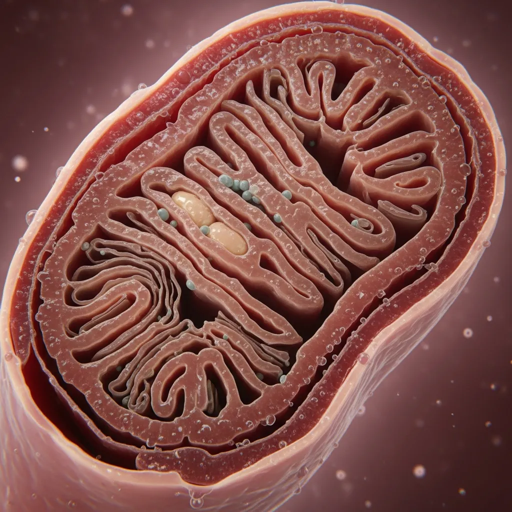
Mitochondrial Biogenesis
Introduction
Your mitochondria are not permanent.
Throughout life, the mitochondrial network inside your cells is continuously being remodeled. Damaged components are repaired or removed, mitochondria divide and fuse, and entirely new mitochondrial material is produced as cells adapt to changes in energy demand.
This process of expanding and rebuilding mitochondrial capacity is known as mitochondrial biogenesis.
The concept sounds simple: make more mitochondria. But the biology is far more sophisticated.
Mitochondria are unusual because they contain their own DNA, yet the overwhelming majority of proteins required to build and operate them are encoded by nuclear DNA. Creating new mitochondrial capacity therefore requires coordination between two separate genomes—the DNA inside the nucleus and the small mitochondrial genome located inside the mitochondria themselves.
At the center of this communication network is a protein called PGC-1α, or peroxisome proliferator-activated receptor gamma coactivator 1-alpha. PGC-1α is frequently described as a "master regulator" of mitochondrial biogenesis because it coordinates numerous transcription factors involved in energy metabolism, oxidative capacity, and mitochondrial adaptation.
But PGC-1α does not become active randomly.
Cells increase mitochondrial capacity when they receive signals that their existing energy-producing machinery is being challenged.
One of the most powerful examples is exercise.
During physical activity, skeletal muscle can increase ATP consumption dramatically. Cellular energy balance changes, calcium concentrations fluctuate, reactive oxygen species temporarily increase, and metabolic sensors such as AMPK respond to the increased energetic demand.
These signals essentially tell the cell:
The current energy-producing capacity is being challenged. We need to adapt.
Repeated exposure to this challenge can stimulate PGC-1α and other regulatory pathways that increase mitochondrial proteins, enzymes involved in oxidative metabolism, and the machinery required to replicate mitochondrial DNA.
Over time, skeletal muscle becomes better equipped to produce ATP through oxidative metabolism.
This is one reason a trained muscle is biologically different from an untrained muscle. Exercise does not simply burn calories while it is being performed. It creates molecular signals that change the machinery responsible for producing energy during future activity.
But mitochondrial biogenesis represents only half of healthy mitochondrial turnover.
In our previous discussion of mitophagy, we explored how cells identify and remove mitochondrial components that have become too dysfunctional to keep. If mitophagy is the quality-control system responsible for removing damaged equipment, mitochondrial biogenesis helps provide the replacement capacity.
The two processes must remain coordinated.
Removing damaged mitochondria without replacing adequate capacity could compromise cellular energy production. Producing new mitochondrial material without eliminating dysfunctional components could allow an increasingly inefficient mitochondrial network to accumulate.
Healthy mitochondrial biology therefore depends on continuous turnover:
Stress → adaptation → repair → removal → recycling → rebuilding.
This balance becomes particularly important in tissues with high energy requirements, including skeletal muscle, the heart, and the brain. Changes in mitochondrial biogenesis have consequently become an important area of research in exercise physiology, insulin resistance, cardiovascular disease, neurodegeneration, muscle aging, and longevity.
The topic has also attracted considerable interest from the supplement and longevity industries. NAD+ boosters, resveratrol, cold exposure, fasting, ketogenic diets, AMPK activators, and numerous other interventions are frequently promoted as ways to "make more mitochondria."
Some influence pathways involved in mitochondrial adaptation. But increasing a molecular marker associated with biogenesis is not the same as demonstrating that an intervention creates healthier mitochondria or improves meaningful human outcomes.
In this article, we'll explore how mitochondrial biogenesis actually works, why PGC-1α plays such an important role, how AMPK and cellular energy stress initiate adaptation, how nuclear and mitochondrial DNA coordinate the construction process, why exercise remains one of the strongest known stimuli, and what mitochondrial biogenesis may teach us about insulin resistance, aging, and metabolic health.
Because mitochondrial health isn't simply about protecting the mitochondria you already have.
Your cells must also know when it's time to build again.
🎧 Listen to the Episode: How Your Body Builds Better Mitochondria
Your cells are constantly adjusting their energy infrastructure to match the demands you place on them. Challenge the system appropriately, recover, and the system adapts. Remove the demand, and that capacity can decline.
In this episode of The Health Pulse, we explore mitochondrial biogenesis, from PGC-1α and TFAM to hormesis, exercise, recovery, and metabolic health. We also separate proven strategies for supporting mitochondrial adaptation from popular biohacks whose promises may be running ahead of the evidence
What Is Mitochondrial Biogenesis?
Mitochondrial biogenesis is the coordinated process through which cells increase and renew their mitochondrial capacity. It involves producing mitochondrial proteins and membranes, replicating mitochondrial DNA, importing proteins from the cytoplasm, and integrating these components into the existing mitochondrial network.
This distinction matters because mitochondria do not simply appear from nothing.
New mitochondrial material is generally produced through the growth and division of existing mitochondria. The process requires extensive coordination between the nucleus, cytoplasm, and mitochondrial network.
The reason is one of the most fascinating features of mitochondrial biology: mitochondria possess their own genome.
Human mitochondrial DNA is a small circular molecule containing approximately 16,500 base pairs. It encodes 13 proteins involved in oxidative phosphorylation, along with 22 transfer RNAs and two ribosomal RNAs required for mitochondrial protein synthesis.
But a functioning mitochondrion requires far more than those 13 proteins.
The vast majority of mitochondrial proteins—well over 1,000—are encoded by nuclear DNA. These include proteins required for nutrient metabolism, mitochondrial dynamics, protein transport, mitochondrial DNA maintenance, antioxidant systems, and many components necessary to assemble and regulate the machinery of oxidative phosphorylation.
Those proteins are produced on cytoplasmic ribosomes and then transported into mitochondria through specialized import systems.
This creates an extraordinary logistical challenge.
To increase mitochondrial capacity, the cell must coordinate gene expression in the nucleus with replication and transcription of mitochondrial DNA. It must manufacture mitochondrial proteins, transport them to the correct location, produce membrane lipids, assemble respiratory-chain complexes, and integrate everything into a functioning mitochondrial network.
This communication between the two genomes is sometimes referred to as mitonuclear coordination.
Several transcriptional regulators help orchestrate the process, but one has become especially important in mitochondrial research: PGC-1α.
PGC-1α does not directly bind DNA like a conventional transcription factor. Instead, it functions as a transcriptional coactivator, interacting with multiple transcription factors and helping increase the expression of genes involved in oxidative metabolism and mitochondrial function.
Among its important partners are nuclear respiratory factors, particularly NRF1 and related transcriptional regulators. These factors stimulate expression of numerous nuclear genes required for mitochondrial function.
One particularly important downstream target is TFAM, or mitochondrial transcription factor A.
TFAM is produced from nuclear DNA, synthesized outside mitochondria, and then imported into the organelle. Once inside, it plays essential roles in packaging mitochondrial DNA and supporting mitochondrial DNA transcription and replication.
This creates a remarkable chain of communication:
Cellular energy demand changes → signaling pathways activate PGC-1α → nuclear mitochondrial genes increase → TFAM is produced → TFAM enters mitochondria → mitochondrial DNA transcription and replication are supported.
The nucleus is therefore helping instruct mitochondria to expand their capacity while the mitochondrial genome simultaneously contributes essential components of the respiratory machinery.
But mitochondrial biogenesis should not be understood simply as increasing the number of mitochondria.
Mitochondria frequently exist as interconnected networks rather than isolated individual organelles. Depending on the tissue and metabolic state, an increase in mitochondrial capacity may involve changes in mitochondrial mass, enzyme content, membrane structure, respiratory proteins, mitochondrial DNA copies, and network organization.
For this reason, scientists often evaluate mitochondrial biogenesis using multiple measurements rather than simply attempting to count mitochondria.
The biological objective is ultimately not to maximize mitochondrial number.
It is to increase the cell's ability to meet its energetic demands efficiently.
A trained skeletal muscle, for example, can develop greater mitochondrial content and oxidative enzyme capacity after repeated endurance exercise. This allows the muscle to produce ATP more effectively through oxidative metabolism, use fatty acids and carbohydrate more efficiently during exercise, and delay reliance on less sustainable energy pathways at a given workload.
Mitochondrial biogenesis is therefore best understood as adaptive remodeling.
The cell experiences repeated energy demand, recognizes that its existing capacity is being challenged, and changes its internal machinery so that the same challenge becomes easier to handle in the future.
Key Takeaway
Mitochondrial biogenesis is not simply the creation of more mitochondria. It is a coordinated remodeling process involving nuclear gene expression, mitochondrial DNA replication, protein production and import, membrane formation, and expansion of oxidative capacity. Because mitochondria depend on genes from both nuclear and mitochondrial DNA, building mitochondrial capacity requires continuous communication between two genomes.
PGC-1α: The Coordinator of Mitochondrial Adaptation
If mitochondrial biogenesis requires hundreds of genes from two different genomes to work together, the cell needs a way to coordinate that response.
One of the most important regulators is PGC-1α, short for peroxisome proliferator-activated receptor gamma coactivator 1-alpha.
PGC-1α is frequently called the “master regulator of mitochondrial biogenesis.” That description is useful, but slightly oversimplified. PGC-1α does not build mitochondria itself, nor does it function as a simple on/off switch. Instead, it acts as a transcriptional coactivator that works with multiple transcription factors to coordinate genes involved in mitochondrial function, oxidative metabolism, fatty acid utilization, and cellular energy adaptation.
This makes PGC-1α particularly important in tissues whose energy requirements can change dramatically, especially skeletal muscle.
During exercise, muscle cells experience several signals simultaneously. ATP is consumed rapidly. AMP and ADP concentrations change. Calcium repeatedly moves through the cell as muscle fibers contract. Reactive oxygen species temporarily increase. Mechanical and metabolic stress activate multiple signaling pathways.
Rather than representing cellular failure, these signals tell the muscle that its existing metabolic machinery has been challenged.
PGC-1α helps translate that challenge into adaptation.
Several pathways can increase PGC-1α activity or expression. One of the best known is AMPK, the cellular energy sensor discussed throughout our previous articles.
When cellular energy availability becomes strained, AMPK becomes activated and helps shift metabolism toward ATP-producing pathways. AMPK can also influence PGC-1α, linking immediate energy stress with longer-term changes in mitochondrial capacity.
Another regulator is SIRT1, an NAD+-dependent deacetylase. SIRT1 can modify PGC-1α and influence its activity, creating a connection between the cellular redox state, NAD+ metabolism, and mitochondrial adaptation.
Muscle contraction introduces another signal through calcium.
Every contraction requires rapid changes in intracellular calcium. Calcium-sensitive pathways—including CaMK signaling—can influence transcriptional programs associated with PGC-1α and oxidative adaptation. This allows repeated muscle contraction itself to become information that eventually changes gene expression.
The result is not one pathway controlling mitochondrial biogenesis, but a network:
Energy stress → AMPK
Redox state → NAD+/SIRT1
Muscle contraction → calcium signaling
Repeated metabolic demand → PGC-1α activation
PGC-1α then interacts with transcription factors including NRF1 and other nuclear regulators that increase expression of genes required for mitochondrial respiration and function.
One important downstream consequence is increased expression of TFAM, the mitochondrial transcription factor discussed in the previous section. TFAM is encoded by nuclear DNA, produced outside the mitochondrion, and transported inside, where it helps regulate mitochondrial DNA maintenance, transcription, and replication.
This creates an elegant biological chain.
The muscle experiences an energetic challenge.
Cellular sensors detect the challenge.
PGC-1α helps coordinate the nuclear response.
Nuclear genes produce proteins needed by mitochondria.
Those proteins are imported into mitochondria.
Mitochondrial DNA and respiratory machinery adapt.
The muscle becomes better prepared for the next challenge.
This is one reason exercise adaptations persist beyond the workout itself.
The calories burned during a training session represent only the immediate energetic cost. The more important long-term effect is that repeated exercise changes the molecular machinery of the muscle.
Over time, endurance training can increase mitochondrial content, oxidative enzymes, capillary density, and the ability of skeletal muscle to generate ATP aerobically. At the same absolute workload, trained muscle can therefore handle the energetic challenge more efficiently than untrained muscle.
PGC-1α also illustrates why mitochondrial health cannot be reduced to taking a compound that activates one pathway.
AMPK, SIRT1, calcium signaling, ROS, PGC-1α, mitochondrial dynamics, mitophagy, and numerous other systems interact continuously. Increasing one molecular signal does not guarantee that the entire mitochondrial network will adapt appropriately.
Exercise is particularly powerful because it activates many of these systems simultaneously and physiologically.
This also helps explain an apparent contradiction in mitochondrial biology.
Exercise temporarily increases energy stress and reactive oxygen species, yet repeated exercise improves mitochondrial function. The temporary stress acts as the signal that tells the cell adaptation is necessary.
The body does not build greater mitochondrial capacity because energy production is always easy.
It builds greater capacity because energy production is repeatedly challenged.
Key Takeaway
PGC-1α helps coordinate the transcriptional response that expands mitochondrial capacity. Signals generated by exercise—including AMPK activation, changes in NAD+-dependent signaling, calcium fluctuations, and metabolic stress—can converge on PGC-1α and related pathways. The result is a coordinated increase in the machinery required for oxidative metabolism and mitochondrial adaptation rather than simply the production of more isolated mitochondria.
Exercise: The Most Powerful Physiological Signal for Mitochondrial Biogenesis
If mitochondrial biogenesis is an adaptation to increased energy demand, exercise provides exactly the type of challenge that should stimulate it.
During physical activity, skeletal muscle can increase ATP consumption dramatically. The existing mitochondrial network must accelerate oxidative phosphorylation to keep pace, while glycolysis, fatty acid oxidation, glycogen breakdown, oxygen delivery, calcium signaling, and numerous other systems adjust simultaneously.
Repeated exercise tells the muscle that its current energy-producing capacity is being challenged on a regular basis.
The long-term response is adaptation.
This is particularly well established with endurance exercise. Repeated aerobic training increases mitochondrial content and oxidative enzyme activity in skeletal muscle, improving the muscle's ability to generate ATP through oxidative metabolism. These adaptations contribute to greater endurance and more efficient fuel utilization at a given workload.
The process begins during the workout itself.
As ATP turnover rises, changes in AMP and ADP contribute to activation of AMPK. Repeated muscle contractions create calcium signals. Cellular redox conditions change, reactive oxygen species temporarily increase, and other kinases respond to mechanical and energetic stress.
These signals converge on transcriptional regulators including PGC-1α.
Importantly, mitochondrial biogenesis does not occur only while someone is exercising. A workout creates the molecular signal, but much of the rebuilding and adaptation occurs during the recovery period that follows.
This distinction is important.
Exercise is the stimulus.
Recovery is when much of the adaptation is constructed.
Repeated cycles of challenge and recovery gradually change skeletal muscle.
Why Endurance Training Has Such a Strong Effect
Activities such as running, cycling, swimming, rowing, and brisk walking require muscles to repeatedly generate ATP over extended periods. Because oxidative phosphorylation is essential for sustaining this activity, endurance training places substantial pressure on the mitochondrial system.
Over time, trained muscle can develop greater mitochondrial density and respiratory capacity, along with increased capillary supply and oxidative enzyme activity.
At the same absolute workload, this means a trained person can often produce a greater proportion of the required ATP aerobically and manage metabolic substrates more effectively.
This is one reason endurance-trained muscle becomes better at using both fat and carbohydrate depending on exercise intensity and fuel availability.
What About Zone 2 Training?
Zone 2 exercise has become particularly popular in discussions of mitochondrial health.
Although definitions vary, Zone 2 generally refers to sustained aerobic exercise performed below the first major lactate threshold, at an intensity where mitochondrial oxidative metabolism can supply much of the required energy and lactate remains relatively controlled.
This type of training can provide a substantial mitochondrial stimulus because it allows relatively large volumes of sustained muscular work without the fatigue associated with very high-intensity exercise.
But there is nothing magical about the term "Zone 2."
Mitochondrial adaptation occurs across a range of exercise intensities, and the ideal program depends on fitness level, health status, training goals, and total exercise volume.
High-Intensity Exercise Also Stimulates Mitochondrial Adaptation
Shorter bouts of high-intensity interval training can also strongly activate pathways associated with mitochondrial biogenesis.
Because high-intensity exercise creates a large energetic disturbance over a relatively short period, signaling through AMPK, calcium-dependent pathways, PGC-1α, and other regulators can be substantial.
Research has shown that interval training can improve mitochondrial content and cardiorespiratory fitness, sometimes with considerably less total training time than traditional endurance programs.
This does not mean high intensity is universally superior.
Higher-intensity exercise creates greater fatigue and may require more recovery. Lower-intensity aerobic work allows greater training volume. A well-designed exercise program can use both.
Resistance Training Matters Too
Resistance exercise is usually associated with muscle hypertrophy and strength, but mitochondrial health should not be separated from muscle mass.
Skeletal muscle represents one of the body's largest metabolically active tissues and an important site of glucose disposal. Maintaining or increasing muscle provides greater capacity for storing glycogen, using glucose, generating force, and remaining physically active.
Resistance training can also influence mitochondrial remodeling, particularly in people who are older, sedentary, or metabolically unhealthy, although its adaptations differ from those produced by endurance training.
This is why reducing mitochondrial health to one type of exercise misses the larger picture.
Aerobic training develops oxidative capacity.
Higher-intensity training challenges maximal metabolic capacity.
Resistance training preserves and builds the tissue containing much of that metabolic machinery.
Together, they create a much more complete stimulus than searching for a single "mitochondrial workout."
The key principle is progressive metabolic demand.
Mitochondria adapt when cells repeatedly encounter an energy requirement that challenges their existing capacity and then receive sufficient recovery to rebuild.
Exercise therefore does something no mitochondrial supplement can currently replicate in its entirety. It simultaneously challenges ATP production, calcium handling, blood flow, glucose transport, fatty acid oxidation, redox signaling, mitochondrial dynamics, mitophagy, and biogenesis.
The result is not merely more mitochondria.
It is a more capable metabolic system.
Key Takeaway
Exercise is one of the strongest established physiological stimuli for mitochondrial biogenesis in humans. Endurance exercise, interval training, and resistance training create different but complementary adaptations. The goal is not to identify one perfect "mitochondrial workout," but to repeatedly challenge cellular energy demand and allow the mitochondrial network to adapt during recovery.
Mitochondrial Biogenesis, Insulin Resistance, and Metabolic Health
The relationship between mitochondria and insulin resistance is often presented as a simple equation:
Fewer or dysfunctional mitochondria → insulin resistance.
The actual biology is considerably more complicated.
People with obesity, type 2 diabetes, physical inactivity, and other metabolic disorders frequently show alterations in skeletal muscle mitochondrial content, oxidative capacity, or mitochondrial gene expression. These observations have led researchers to investigate whether impaired mitochondrial function contributes directly to insulin resistance.
There is good reason for the interest.
Skeletal muscle is one of the body's largest sites of insulin-stimulated glucose disposal. After a carbohydrate-containing meal, insulin helps move glucose into muscle cells, where it can be stored as glycogen or oxidized for energy.
A metabolically active muscle with substantial mitochondrial capacity can handle enormous amounts of fuel.
Problems can emerge when energy availability chronically exceeds the amount being used.
In an inactive individual consuming more energy than tissues require, fatty acids and other metabolic intermediates can accumulate within skeletal muscle. Certain lipid intermediates, particularly diacylglycerols and ceramides, have been implicated in signaling pathways that interfere with normal insulin action.
This creates an important distinction.
The presence of fat inside skeletal muscle is not automatically pathological.
Endurance athletes can store considerable amounts of intramuscular triglyceride while remaining highly insulin sensitive—a phenomenon sometimes called the athlete's paradox. The difference appears to involve factors such as mitochondrial oxidative capacity, lipid turnover, cellular localization of lipid species, physical activity, and the ability to rapidly use stored fuel.
In other words, metabolic health depends not simply on how much fuel enters a cell, but on the cell's capacity to process, store, mobilize, and oxidize that fuel appropriately.
Mitochondrial biogenesis can contribute to that capacity.
Exercise-induced increases in mitochondrial enzymes and oxidative machinery allow skeletal muscle to oxidize more substrate when energy demand rises. Exercise also increases glucose transport through mechanisms that do not depend entirely on insulin, while repeated training improves insulin sensitivity over time.
However, this does not mean that increasing mitochondrial number alone will automatically reverse insulin resistance.
Which Comes First?
This remains an important scientific question.
Does mitochondrial dysfunction cause insulin resistance?
Or does insulin resistance, chronic energy excess, physical inactivity, inflammation, and altered substrate availability eventually impair mitochondrial function?
Evidence suggests the relationship can operate in both directions, and the answer likely differs according to the individual and metabolic context.
For example, physical inactivity can reduce mitochondrial oxidative capacity because muscle no longer requires the same energy-producing machinery. This may be an adaptation to reduced demand rather than a primary mitochondrial disease.
At the same time, chronic nutrient excess and lipid accumulation can create metabolic stress that alters mitochondrial function and insulin signaling.
Genetics, aging, sleep, hormones, adipose tissue function, liver metabolism, muscle mass, and physical activity all contribute to the larger system.
This is why insulin resistance should not be reduced to a mitochondrial problem alone.
Adipose Tissue Still Matters
Skeletal muscle mitochondria are only one part of whole-body insulin sensitivity.
Healthy adipose tissue acts as a storage buffer, safely retaining large amounts of energy as triglycerides. As adipose tissue becomes dysfunctional or reaches an individual's storage capacity, more fatty acids may spill into the circulation and reach tissues such as the liver and skeletal muscle.
This ectopic fuel delivery can contribute to fatty liver, increased VLDL production, lipid accumulation within muscle, and impaired insulin signaling.
The liver simultaneously plays a central role in controlling glucose production and lipid metabolism.
Insulin resistance is therefore better understood as a disturbance involving adipose tissue, skeletal muscle, liver, pancreas, and cellular energy metabolism together.
Mitochondrial biogenesis matters because it can increase the ability of metabolically active tissues—especially skeletal muscle—to respond to energy demand.
But creating additional oxidative capacity is most useful when that capacity is actually used.
A person cannot simply build mitochondria and expect them to continuously burn excess energy while remaining sedentary. Mitochondrial metabolism responds strongly to demand.
This brings us back to exercise.
Exercise simultaneously increases energy expenditure, stimulates mitochondrial biogenesis, improves insulin-independent glucose uptake during contraction, improves subsequent insulin sensitivity, preserves or builds skeletal muscle, and increases the metabolic demand placed on that tissue.
Rather than targeting one molecular defect, it changes the entire metabolic environment.
That may be one reason exercise remains such a powerful intervention for insulin resistance.
Key Takeaway
Mitochondrial dysfunction and insulin resistance are closely associated, but the relationship is not simply cause and effect. Metabolic health depends on the ability of skeletal muscle, adipose tissue, and the liver to appropriately store, mobilize, and use energy. Exercise-induced mitochondrial biogenesis can increase skeletal muscle oxidative capacity and improve insulin sensitivity, but increasing mitochondrial number alone is not a cure for insulin resistance.
Aging: Why Mitochondrial Renewal Becomes More Important With Time
Aging is often described as a gradual loss of cellular function. Mitochondria sit near the center of this process because tissues must continuously maintain the organelles responsible for producing much of their usable energy.
As we age, mitochondrial biology changes.
Mitochondrial DNA can accumulate mutations and damage, respiratory-chain function may become less efficient in some tissues, mitochondrial dynamics can change, and the systems responsible for identifying and removing dysfunctional mitochondria may become less effective. At the same time, mitochondrial biogenesis and the signaling pathways that coordinate adaptation may become less responsive.
The result is not simply "fewer mitochondria."
It can be a decline in mitochondrial turnover and adaptability.
This distinction is important because mitochondrial health depends on both sides of the quality-control equation. As we discussed in the previous article, mitophagy helps remove mitochondrial components that are too damaged to retain. Mitochondrial biogenesis helps restore and expand functional capacity.
Healthy aging therefore requires coordination between:
Removing what no longer works → preserving what still works → rebuilding what has been lost.
If removal slows while replacement also becomes less responsive, dysfunctional mitochondrial components can gradually accumulate.
Skeletal Muscle Makes the Problem More Important
One of the most visible consequences of aging is the progressive decline in skeletal muscle mass, strength, and physical capacity.
Muscle loss is not purely a mitochondrial problem. Changes in physical activity, motor neurons, hormones, protein intake, inflammation, anabolic signaling, illness, and other factors contribute.
But declining mitochondrial function can make muscle less capable of meeting energetic demands, while declining muscle mass reduces the amount of metabolically active tissue available to use glucose and fatty acids.
This can create a damaging feedback loop.
A person becomes less active.
Lower activity reduces the metabolic demand placed on skeletal muscle.
Reduced demand provides less stimulus for mitochondrial adaptation.
Physical capacity declines further.
Activity becomes increasingly difficult.
Over years, this cycle can contribute to worsening metabolic flexibility, insulin resistance, frailty, and loss of independence.
This is one reason preserving physical capacity with age matters far beyond appearance or athletic performance.
Exercise Can Still Stimulate Aging Mitochondria
One of the most encouraging findings from exercise physiology is that older skeletal muscle retains a substantial capacity to adapt.
Aerobic exercise can increase mitochondrial proteins, oxidative enzymes, and respiratory capacity in older adults. Resistance training can increase strength and preserve or restore muscle mass, while also improving metabolic function.
The mitochondrial response may differ from that of a younger person, and factors such as training history, disease, nutrition, and recovery matter. But aging does not eliminate the ability to generate meaningful mitochondrial adaptations.
This has major implications.
Rather than viewing age-related mitochondrial decline as an unavoidable one-way process, physical activity can continue providing signals that tell older muscle:
This energy-producing machinery is still required. Maintain it.
Mitochondrial Biogenesis and Longevity
Researchers have naturally asked whether increasing mitochondrial biogenesis could extend lifespan.
The answer is not yet clear.
Long-lived organisms frequently demonstrate effective stress-response systems, metabolic flexibility, protein quality control, autophagy, and mitochondrial maintenance. Pathways involving AMPK, sirtuins, NAD+, mTOR, PGC-1α, and mitophagy repeatedly appear in aging research.
But longevity cannot be reduced to maximizing any one of them.
More mitochondrial biogenesis is not automatically better, just as more mitophagy is not automatically better. Producing additional mitochondria without appropriate quality control could simply expand a dysfunctional network.
The more meaningful concept may be mitochondrial turnover—maintaining the ability to remove damaged components and generate functional replacements as conditions change.
This is where exercise again becomes especially interesting.
Exercise does not target only mitochondrial biogenesis. It simultaneously challenges energy production, stimulates mitochondrial remodeling, supports mitophagy, improves cardiovascular fitness, preserves skeletal muscle, improves insulin sensitivity, and activates multiple cellular stress-response pathways.
These adaptations may help explain why measures of physical fitness—particularly cardiorespiratory fitness—are so strongly associated with long-term health outcomes.
That does not prove that mitochondrial biogenesis itself extends human lifespan.
It suggests something more practical: maintaining the body's capacity to respond and adapt to energetic stress appears to be an important feature of healthy aging.
The objective is therefore not to preserve mitochondria in exactly the state they existed when we were young.
Healthy aging requires mitochondria that can continue to change.
Key Takeaway
Aging can impair mitochondrial quality control, turnover, and responsiveness, particularly as physical activity and skeletal muscle decline. However, older muscle retains a meaningful capacity for mitochondrial adaptation. Regular aerobic and resistance exercise can continue stimulating mitochondrial remodeling while preserving the metabolically active tissue needed for glucose disposal, physical function, and healthy aging.
Can Fasting, Ketosis, or Cold Exposure Build More Mitochondria?
Exercise has some of the strongest human evidence for stimulating mitochondrial adaptation, but it is not the only physiological stress capable of influencing pathways involved in mitochondrial biogenesis.
Fasting, carbohydrate restriction, ketosis, and cold exposure all change the energetic environment of the cell. Because mitochondrial biogenesis is partly regulated by energy sensing, researchers have investigated whether these interventions can increase mitochondrial capacity.
The answer is interesting—but much more nuanced than simply saying they "make more mitochondria."
Fasting and Energy Sensing
During fasting, insulin levels generally fall while the body shifts toward greater reliance on stored energy. Liver glycogen declines, adipose tissue releases more fatty acids, and the liver can increase production of ketone bodies as fasting progresses.
At the cellular level, reduced nutrient availability can influence AMPK, mTOR, sirtuins, NAD+ metabolism, autophagy, and PGC-1α-associated pathways.
This creates a biological environment compatible with mitochondrial remodeling.
Fasting may therefore influence both sides of mitochondrial turnover: pathways involved in removing damaged cellular material and pathways involved in adapting energy metabolism to changing fuel availability.
But there is no universal fasting duration that has been proven to maximize mitochondrial biogenesis in humans.
Claims that mitochondrial biogenesis suddenly begins after 16, 24, 48, or 72 hours of fasting oversimplify a dynamic process that differs among tissues and individuals.
Longer fasting is also not automatically better. Extended fasting can reduce training performance and, depending on duration, frequency, protein intake, and individual circumstances, contribute to loss of lean tissue.
For mitochondrial health, preserving skeletal muscle remains extremely important.
Ketosis and Mitochondrial Adaptation
Ketosis creates another interesting metabolic environment.
When carbohydrate availability becomes sufficiently low, the liver converts fatty acids into ketone bodies, primarily beta-hydroxybutyrate (BHB) and acetoacetate. These molecules can be transported to tissues and oxidized within mitochondria for energy.
BHB is also more than a fuel molecule.
Research suggests that beta-hydroxybutyrate can participate in cellular signaling, influence gene expression, alter redox biology, and interact with pathways involved in oxidative stress and metabolic adaptation.
Animal studies and experimental models have reported changes in mitochondrial biogenesis and mitochondrial function during ketogenic interventions in certain tissues.
However, the human evidence is more complicated.
A ketogenic diet does not automatically produce more mitochondria simply because ketones are present. The mitochondrial response depends on energy demand, tissue type, physical activity, calorie balance, training status, and metabolic health.
A sedentary person in nutritional ketosis does not necessarily receive the same mitochondrial stimulus as someone repeatedly challenging skeletal muscle through exercise.
This distinction is essential.
Changing the fuel is not the same as increasing the demand for energy production.
Ketogenic diets may provide therapeutic or metabolic benefits in selected circumstances, and their effects on mitochondrial signaling remain an active area of research. But they should not be described as a substitute for exercise-induced mitochondrial adaptation.
Cold Exposure and Brown Fat
Cold exposure provides an entirely different mitochondrial challenge.
When the body is exposed to cold, maintaining core temperature requires increased heat production. One tissue particularly involved in this response is brown adipose tissue.
Unlike white adipose tissue, which primarily stores energy, brown adipose tissue contains large numbers of mitochondria and specializes in thermogenesis.
A mitochondrial protein called uncoupling protein 1 (UCP1) allows brown-fat mitochondria to dissipate part of the proton gradient as heat rather than capturing all of that energy as ATP.
Repeated cold exposure can activate sympathetic signaling and increase thermogenic activity. Experimental and human research suggests that cold acclimation can influence brown adipose tissue quantity, mitochondrial activity, and glucose metabolism.
This makes cold exposure an interesting example of mitochondrial adaptation occurring for a purpose other than muscle contraction.
The mitochondria are responding to increased thermoregulatory demand.
But once again, this does not mean increasingly extreme cold produces increasingly better mitochondrial health.
Cold plunges and cryotherapy have become popular wellness interventions, yet much of the mechanistic enthusiasm exceeds evidence for long-term clinical benefits. Cold exposure also creates cardiovascular stress and is not appropriate for everyone.
Hormesis: The Common Thread
Exercise, fasting, ketosis, and cold exposure appear very different, but they share an important biological principle.
Each can create a temporary challenge to cellular homeostasis.
This concept is often described as hormesis: a manageable stressor activates adaptive responses that can make the organism better prepared for future stress.
But hormesis has an important limitation.
A stressor is beneficial only when the organism can recover and adapt.
Too little stress may produce little adaptation.
An appropriate challenge may improve resilience.
Excessive stress can overwhelm the system and cause damage.
This is why mitochondrial health should not become a competition to accumulate as many stressors as possible through prolonged fasting, extreme exercise, severe carbohydrate restriction, cold plunges, heat exposure, and other interventions simultaneously.
The objective is adaptation—not suffering.
Among these interventions, exercise remains the most consistently supported strategy for increasing skeletal muscle mitochondrial capacity in humans. Fasting, ketosis, and cold exposure influence interesting metabolic pathways and may have specific applications, but their effects should be understood within the broader context of energy demand, recovery, nutrition, and individual health.
Key Takeaway
Fasting, ketosis, and cold exposure can influence energy-sensing and mitochondrial pathways, but none should be viewed as a simple switch for mitochondrial biogenesis. Their effects depend on tissue, duration, metabolic state, and energy demand. Exercise remains the best-established physiological stimulus for increasing skeletal muscle mitochondrial capacity in humans.
Can Supplements Increase Mitochondrial Biogenesis?
The idea of building more mitochondria has created an enormous market for supplements marketed as “mitochondrial boosters.” NAD+ precursors, resveratrol, CoQ10, PQQ, creatine, carnitine, alpha-lipoic acid, and numerous other compounds are promoted with claims ranging from increased energy to improved longevity.
Some of these compounds participate in pathways related to mitochondrial biology. But that does not mean taking them automatically causes meaningful mitochondrial biogenesis in humans.
This distinction is essential.
NAD+ Precursors
NAD+ is a central molecule in cellular energy metabolism. It participates in redox reactions that allow nutrients to be converted into usable energy and also serves as a substrate for enzymes including sirtuins and PARPs.
Because SIRT1 can influence PGC-1α, researchers have investigated whether increasing NAD+ availability could enhance mitochondrial adaptation.
This has driven interest in NAD+ precursors such as nicotinamide riboside (NR) and nicotinamide mononucleotide (NMN).
Human trials show that these compounds can increase NAD+-related metabolites in blood and sometimes in tissues. That establishes an important biological effect.
But raising NAD+ is not the same as demonstrating improved mitochondrial function.
Clinical trials have produced mixed results for outcomes such as insulin sensitivity, muscle performance, exercise capacity, and other measures of metabolic health. Some studies report potentially beneficial signals in selected populations, while others show little meaningful physiological improvement despite successful increases in NAD+ availability.
This is a recurring lesson in mitochondrial research:
Changing a biomarker does not necessarily change an outcome.
Resveratrol
Resveratrol became popular partly because experimental research connected it with SIRT1, AMPK, and PGC-1α-associated signaling.
Animal and cellular experiments generated considerable excitement around the possibility that resveratrol might mimic some effects of calorie restriction or exercise.
Human evidence has been far less dramatic.
Studies have reported inconsistent effects on metabolic health, mitochondrial function, and exercise adaptation. Importantly, some research has even raised the possibility that high-dose antioxidant-like supplementation could blunt portions of the normal adaptive response to exercise under certain circumstances.
This makes biological sense.
As discussed throughout this series, temporary energetic and oxidative stress helps tell the cell that adaptation is required. Eliminating every stress signal may therefore interfere with some of the communication responsible for adaptation.
Coenzyme Q10
Coenzyme Q10 (CoQ10) occupies an important position within the mitochondrial electron transport chain, transferring electrons between respiratory complexes.
CoQ10 is therefore undeniably important for mitochondrial function.
But an essential molecule is not automatically beneficial when supplemented in unlimited amounts.
CoQ10 supplementation has established and investigational uses in particular clinical contexts, and some people may benefit depending on their health status or medications. However, taking CoQ10 does not necessarily stimulate mitochondrial biogenesis in a healthy person with adequate levels.
Supporting mitochondrial function and building additional mitochondrial capacity are not the same biological process.
PQQ
Pyrroloquinoline quinone (PQQ) is frequently marketed specifically as a mitochondrial biogenesis supplement.
Some cell and animal studies have reported effects involving PGC-1α and pathways associated with mitochondrial formation. These findings are biologically interesting.
However, strong human evidence demonstrating that PQQ supplementation meaningfully increases mitochondrial content and improves clinically important outcomes remains limited.
Marketing claims have therefore moved considerably faster than the clinical science.
Creatine
Creatine deserves a slightly different discussion.
Creatine does not primarily work by stimulating mitochondrial biogenesis. Instead, the phosphocreatine system helps rapidly buffer and regenerate ATP during periods of high energy demand.
This makes creatine particularly useful for skeletal muscle performance, strength, and training capacity, with a much larger body of human evidence than many supplements marketed specifically for mitochondrial health.
Creatine may indirectly support mitochondrial health by allowing people to train more effectively, maintain skeletal muscle, and repeatedly create the energetic stimulus that drives adaptation.
That distinction is useful.
Sometimes the best way to support mitochondrial biology is not to directly target mitochondria with a supplement.
It is to improve the body's ability to perform the activity that stimulates mitochondrial adaptation naturally.
Nutrient Deficiency Is Different
There is one situation where supplementation can make a substantial difference: correcting a genuine deficiency.
Mitochondrial metabolism depends on numerous vitamins and minerals, including iron, several B vitamins, magnesium, and other micronutrients that serve as cofactors in energy-producing pathways.
If someone is deficient in an essential nutrient, restoring adequate status may improve normal physiological function.
But correcting deficiency is fundamentally different from taking supraphysiological amounts of nutrients in an attempt to "supercharge" mitochondria.
Once nutritional requirements are met, more is not necessarily better.
This is why targeted laboratory testing can sometimes be more useful than blindly assembling a large mitochondrial supplement stack.
The Exercise Comparison
There is also a practical benchmark against which mitochondrial supplements should be evaluated.
Can the intervention reproduce what exercise does?
Exercise changes ATP turnover, calcium signaling, AMPK activity, PGC-1α signaling, glucose transport, fatty acid oxidation, mitochondrial dynamics, mitophagy, vascular function, cardiorespiratory fitness, and skeletal muscle simultaneously.
No currently available supplement has been demonstrated to reproduce this entire physiological response.
That does not mean mitochondrial supplements are useless. Some may have legitimate applications, and several remain interesting areas of research.
It simply means the evidence should match the claim.
A supplement that increases NAD+ metabolites or changes PGC-1α expression in an experimental model should not automatically be advertised as though it has been proven to create younger mitochondria, reverse metabolic disease, or extend human lifespan.
Mitochondrial biogenesis is a coordinated adaptation to demand.
And at present, one of the most reliable ways to create that demand remains remarkably simple:
Use the mitochondria you want your body to maintain.
Key Takeaway
Several supplements influence pathways related to mitochondrial biology, but evidence that they meaningfully increase mitochondrial capacity or improve clinical outcomes in humans varies considerably. Correcting genuine nutrient deficiencies can support normal energy metabolism, while exercise remains the best-established way to create the physiological demand that drives skeletal muscle mitochondrial adaptation.
How Lab Testing Fits Into Mitochondrial Biogenesis
After learning about PGC-1α, AMPK, NAD+, mitophagy, and mitochondrial biogenesis, it is tempting to look for a blood test that can answer a seemingly simple question:
How well are my mitochondria working?
For routine metabolic health, there is currently no single blood test that can reliably answer that question.
Researchers can measure mitochondrial DNA copy number, respiratory capacity, oxidative enzymes, gene expression, PGC-1α signaling, and other aspects of mitochondrial biology. Specialized testing can also evaluate oxygen consumption and ATP-related processes in cells or tissue samples.
These techniques are extremely useful in research and in selected clinical situations involving suspected mitochondrial disorders. But they should not be confused with a validated routine “mitochondrial health panel.”
There is also no established blood concentration of PGC-1α, AMPK, NAD+, or another single marker that can tell someone how many healthy mitochondria they have or whether they need to increase mitochondrial biogenesis.
For most people, laboratory testing is more useful for answering a different question:
Is there something measurable interfering with normal energy metabolism?
Insulin and Glucose Regulation
One of the most useful places to begin is glucose metabolism.
Fasting glucose and HbA1c provide information about blood glucose regulation, while fasting insulin can add context about the amount of insulin required to maintain that glucose.
This distinction matters because glucose can remain within the normal range while insulin concentrations are already elevated. Compensatory hyperinsulinemia may therefore provide an earlier clue that skeletal muscle, adipose tissue, or the liver is becoming less responsive to insulin.
Because skeletal muscle is a major site of glucose disposal and mitochondrial adaptation, identifying insulin resistance provides useful information about the metabolic environment in which muscle mitochondria are functioning.
Lipids and Liver Metabolism
A standard lipid panel provides another window into energy handling.
Elevated triglycerides, particularly when accompanied by low HDL cholesterol and other features of metabolic syndrome, may suggest increased hepatic VLDL production and insulin resistance.
Advanced cardiovascular markers such as ApoB can help quantify atherogenic particle burden, while liver-related markers including ALT and AST may provide additional context.
However, normal liver enzymes do not exclude metabolic dysfunction-associated steatotic liver disease. When fatty liver is suspected, additional clinical evaluation or imaging may be appropriate.
Oxygen Delivery Matters
Mitochondria cannot perform oxidative phosphorylation without oxygen.
That means problems outside the mitochondria can dramatically affect what feels like an energy-production problem.
A complete blood count (CBC) can help identify anemia and abnormalities involving red blood cells or hemoglobin. Ferritin and iron studies may reveal iron deficiency, which can impair oxygen delivery and also affect iron-dependent proteins involved in mitochondrial metabolism.
Someone experiencing fatigue or poor exercise tolerance because of iron deficiency does not necessarily need a mitochondrial supplement.
They may need the underlying deficiency identified and appropriately treated.
Thyroid Function
Thyroid hormones strongly influence metabolic rate, oxygen consumption, thermogenesis, and mitochondrial activity.
Testing TSH and free T4, with additional thyroid evaluation when clinically appropriate, can help identify thyroid dysfunction that may contribute to fatigue, altered lipid metabolism, temperature intolerance, or changes in energy expenditure.
Again, these symptoms can easily be attributed online to "poor mitochondria" despite having a completely different—and measurable—cause.
Nutrient Status
Mitochondrial energy pathways require numerous micronutrients as cofactors.
Depending on diet, symptoms, medications, and medical history, testing may include vitamin B12, folate, ferritin, iron studies, vitamin D, magnesium, and other targeted nutrients.
The objective should not be to push every nutrient toward the highest possible laboratory value.
It is to identify genuine deficiencies or abnormalities that could interfere with normal physiology.
Inflammation and Cardiovascular Risk
High-sensitivity C-reactive protein (hs-CRP) can provide information about systemic inflammation and cardiovascular risk. It does not measure mitochondrial inflammation or mitochondrial dysfunction specifically, but persistent metabolic inflammation can exist alongside insulin resistance and other conditions affecting cellular energy metabolism.
Advanced cardiovascular testing may also include ApoB, lipoprotein(a), and additional lipoprotein measurements when appropriate.
This broader perspective matters because mitochondrial health cannot be separated from vascular health.
Mitochondria need oxygen and nutrients delivered through a functioning cardiovascular system. Muscle needs adequate blood flow. The heart itself depends heavily on mitochondrial ATP production.
Energy metabolism is a whole-body system.
When Specialized Mitochondrial Testing Is Appropriate
True mitochondrial diseases are different from the vague concept of "suboptimal mitochondrial health."
Inherited mitochondrial disorders can produce neurological symptoms, muscle weakness, exercise intolerance, developmental problems, hearing or vision abnormalities, seizures, cardiomyopathy, and other potentially serious manifestations.
Evaluation may involve lactate, creatine kinase, metabolic studies, genetic testing, tissue analysis, or specialized measurements of mitochondrial function, depending on the clinical presentation.
These investigations belong within specialized medical evaluation and should not be marketed as general longevity screening.
At QuickLab Mobile, we provide convenient at-home laboratory testing throughout Miami for many of the metabolic factors surrounding cellular energy health, including fasting insulin, glucose, HbA1c, lipid testing, ApoB, hs-CRP, CBC, ferritin and iron studies, thyroid markers, vitamin B12, vitamin D, and comprehensive metabolic testing.
These tests cannot count your mitochondria or determine whether your PGC-1α is "optimized."
What they can do is identify measurable abnormalities that may be affecting the environment in which your mitochondria are expected to perform.
That is often a far more useful place to begin.
Key Takeaway
There is no routine blood test that directly measures mitochondrial biogenesis or provides a reliable mitochondrial health score. Laboratory testing is more useful for identifying insulin resistance, anemia, iron or nutrient deficiencies, thyroid dysfunction, inflammation, liver abnormalities, and other measurable factors that can affect energy metabolism and physical performance.
Conclusion
Mitochondria are not static structures that we are born with and slowly lose over time. They belong to a dynamic cellular network that is continually being remodeled in response to how much energy our tissues require.
Mitochondrial biogenesis is one of the mechanisms that makes this adaptation possible.
When cells repeatedly encounter increased energy demand, molecular signals communicate that existing capacity is being challenged. AMPK responds to cellular energy stress, calcium signaling reflects repeated muscle contraction, NAD+-dependent pathways interact with metabolic regulators, and PGC-1α helps coordinate the transcriptional response.
The result is far more sophisticated than simply producing additional mitochondria.
Nuclear DNA must increase expression of mitochondrial proteins. Those proteins must be manufactured and transported into mitochondria. TFAM helps coordinate mitochondrial DNA maintenance and expression. Membranes and respiratory-chain components must be assembled, and the resulting mitochondrial material must integrate into an already dynamic network.
Two genomes effectively cooperate to expand the cell's capacity to produce energy.
But mitochondrial biogenesis is only useful when paired with mitochondrial quality control.
As we explored in our discussion of mitophagy, cells must continually identify mitochondrial components that are damaged beyond repair and remove them. Fission and fusion remodel the network, mitophagy eliminates dysfunctional material, and mitochondrial biogenesis restores capacity.
Together, these processes create continuous mitochondrial turnover:
Challenge → adaptation → quality control → removal → rebuilding.
This is why simply trying to maximize mitochondrial number misses the point.
A cell containing large numbers of poorly functioning mitochondria is not necessarily healthier. Likewise, constantly stimulating mitophagy without adequately replacing mitochondrial capacity would not be desirable.
The goal is a mitochondrial network capable of adapting to changing energy demands.
Exercise remains one of the clearest demonstrations of this principle.
Physical activity temporarily disrupts energy balance. ATP demand rises, calcium signaling changes, oxidative signals increase, and existing mitochondrial capacity is challenged. Rather than being inherently harmful, these temporary stresses become instructions for adaptation.
During recovery, the body rebuilds.
Repeated over months and years, this cycle can increase skeletal muscle oxidative capacity, improve insulin sensitivity, support cardiovascular fitness, and preserve physical function.
Fasting, ketosis, cold exposure, NAD+ precursors, and other interventions may influence some of the same signaling pathways, and several remain valuable areas of research. But none should distract from the larger biological principle.
Mitochondria adapt to demand.
This becomes especially important as we age.
Maintaining skeletal muscle, aerobic capacity, and regular physical activity continues sending a powerful signal that mitochondrial capacity is necessary. Rather than allowing declining activity to progressively reduce metabolic demand, exercise gives aging tissues a reason to preserve and rebuild their energy-producing machinery.
Laboratory testing cannot currently tell us how many healthy mitochondria someone has or provide a simple mitochondrial biogenesis score. But it can identify abnormalities in glucose regulation, insulin, thyroid function, iron status, nutrient availability, inflammation, liver health, and cardiovascular risk that may affect the larger metabolic environment.
At QuickLab Mobile, we provide convenient at-home laboratory testing throughout Miami for patients who want objective information about these measurable components of metabolic health.
Understanding mitochondrial biology ultimately changes the way we think about energy.
Your cells do not simply possess a fixed amount of energy-producing machinery.
They are constantly asking a question:
How much capacity does this body actually require?
How we move, train, recover, eat, and age helps provide the answer.
Metabolic health involves much more than mitochondrial number. QuickLab Mobile offers convenient at-home testing throughout Miami for fasting insulin, glucose, HbA1c, advanced cardiovascular markers, thyroid function, nutrient status, inflammation, and other biomarkers that can help reveal the metabolic environment affecting cellular energy production.
👉 Need a specimen collection?. Book Now
Disclaimer:
The information provided in this blog, podcast, and associated content is for educational and informational purposes only and is not intended as a substitute for professional medical advice, diagnosis, or treatment. The content shared is based on reputable sources, medical literature, and expert insights, but it should not be used as a replacement for direct consultation with a licensed healthcare provider.
No Doctor-Patient Relationship: Engaging with this content does not create a doctor-patient relationship between you and QuickLabMobile or any contributors. Always consult with a qualified physician, specialist, or healthcare professional before making any medical decisions, changing your treatment plan, or starting/stopping any medications.
Not a Substitute for Medical Advice: While we strive to provide accurate and up-to-date information, medicine is constantly evolving. New research, treatments, and medical recommendations may emerge, and individual health conditions can vary. Do not rely solely on this content for health decisions. If you are experiencing symptoms, have concerns about your health, or require medical assistance, seek immediate care from a licensed medical professional.
Emergency Situations: If you are experiencing a medical emergency, such as difficulty breathing, chest pain, signs of a stroke, or any other life-threatening condition, call 911 (or your local emergency services) immediately. Do not delay seeking emergency care based on information provided here.
Liability Disclaimer: QuickLabMobile, its contributors, and any associated entities do not assume liability for any damages, harm, or adverse outcomes resulting from the use, interpretation, or misuse of the information provided in this content. You are responsible for your own healthcare decisions and should always verify information with a trusted medical professional.
External Links & References: This content may include links to external sources, medical studies, or third-party websites for further reading. These links are provided for convenience and informational purposes only. QuickLabMobile does not endorse, control, or take responsibility for the accuracy of external content. Always verify information with authoritative sources such as the CDC, NIH, WHO.
Final Note: Your health is unique, and what works for one person may not be suitable for another. Stay informed, ask questions, and always prioritize professional medical guidance