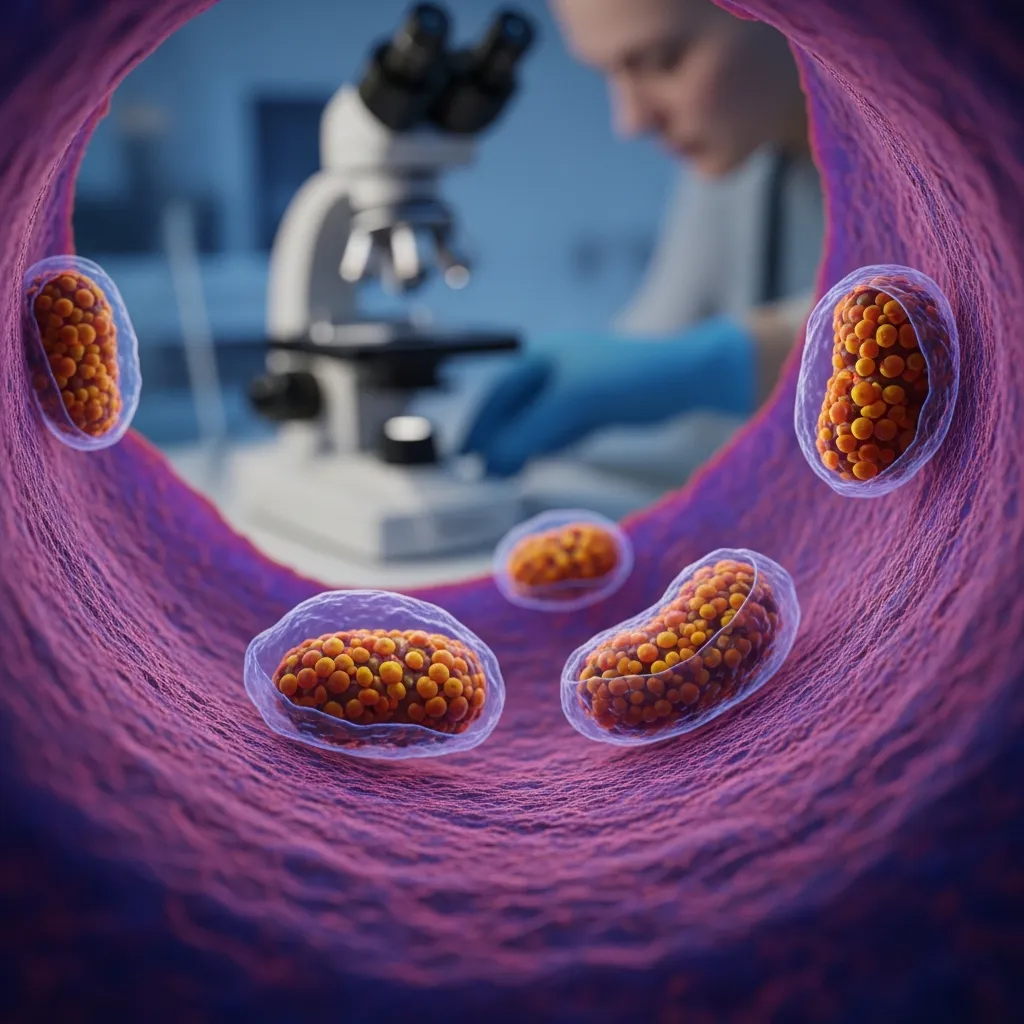
Mitophagy: How Your Cells Find and Remove Damaged Mitochondria
Introduction
Mitochondria are constantly under stress.
Every second, these tiny organelles convert nutrients into ATP through oxidative phosphorylation, supplying the energy required for muscle contraction, brain activity, cellular repair, ion transport, and virtually every other energy-dependent process in the body. But producing energy comes with a cost.
Mitochondria are continuously exposed to reactive oxygen species, changes in membrane potential, damaged proteins, mutations in mitochondrial DNA, and fluctuations in nutrient and energy availability. Over time, some mitochondria inevitably become dysfunctional.
The cell therefore faces an important problem: what should it do with a mitochondrion that is too damaged to function properly?
The answer is mitophagy.
Mitophagy is a specialized form of autophagy through which cells identify damaged or unnecessary mitochondria, isolate them, and deliver their components to lysosomes for degradation and recycling. Rather than allowing dysfunctional mitochondria to accumulate indefinitely, the cell operates a sophisticated mitochondrial quality-control system that continually evaluates which mitochondria should remain and which should be removed.
This process is essential because a severely dysfunctional mitochondrion can become more than an inefficient energy producer. Loss of mitochondrial membrane potential, impaired electron transport, excessive reactive oxygen species production, abnormal calcium handling, and release of mitochondrial components can create cellular stress and activate inflammatory or apoptotic pathways.
Removing damaged mitochondria therefore helps protect the entire mitochondrial network.
But destruction is only half of the story.
Healthy mitochondrial function depends on a continuous cycle of damage, detection, removal, and replacement. Damaged mitochondria can be separated from healthier portions of the mitochondrial network through mitochondrial fission. Mitophagy then removes components that cannot be adequately repaired, while mitochondrial biogenesis helps generate new mitochondrial material.
Together, these processes create something resembling cellular quality control.
This becomes particularly important in tissues with enormous energy requirements. Skeletal muscle, the heart, and the brain depend heavily on mitochondrial ATP production and must maintain functioning mitochondrial populations throughout life. When mitochondrial quality-control mechanisms begin to fail, dysfunctional mitochondria may accumulate and contribute to metabolic dysfunction, muscle decline, neurodegeneration, inflammation, and other features associated with aging.
Mitophagy has therefore become an important area of research in conditions ranging from insulin resistance and cardiovascular disease to Parkinson's disease and age-related muscle loss.
The growing interest in mitophagy has also produced considerable enthusiasm around fasting, exercise, ketogenic diets, supplements, and compounds claimed to "activate autophagy." Some of these interventions influence pathways involved in cellular quality control, but the human evidence is considerably more nuanced than many online claims suggest.
In this article, we'll explore how cells recognize damaged mitochondria, the remarkable PINK1-Parkin pathway, the relationship between mitochondrial fission and mitophagy, how exercise and nutrient availability influence mitochondrial turnover, what happens when mitophagy fails, and what current research actually tells us about improving mitochondrial quality.
Because when it comes to mitochondrial health, producing more mitochondria is only part of the equation.
Sometimes the healthiest thing a cell can do is destroy the mitochondria it already has.
🎧 Listen to the Episode: Your Mitochondria Need a Cleanup System
More mitochondria aren't necessarily better. What matters is whether the mitochondria you have can efficiently produce energy without becoming a source of excessive oxidative stress and inflammation.
In this episode of The Health Pulse, we explore mitophagy—the cellular recycling system responsible for identifying and removing damaged mitochondria. From PINK1 and Parkin to exercise, AMPK, mitohormesis, and mitochondrial biogenesis, we break down how the body maintains its cellular power grid and what happens when that quality-control system begins to fail.
▶️ Click play below to listen, or keep reading to discover why improving cellular energy may begin not by adding more—but by clearing away what no longer works.
What Is Mitophagy?
To understand mitophagy, we first need to understand autophagy.
Autophagy literally means "self-eating." Despite the dramatic name, it is a normal and essential cellular recycling system. Cells constantly accumulate damaged proteins, worn-out structures, and other material that must either be repaired or removed. Autophagy allows cells to package some of this material and deliver it to lysosomes, specialized compartments containing enzymes capable of breaking cellular components into reusable building blocks.
Mitophagy is the mitochondrial version of this quality-control process.
Rather than randomly destroying mitochondria, cells possess mechanisms that help identify mitochondria that have become damaged, dysfunctional, or unnecessary. Selected mitochondria can then be enclosed within an autophagosome, a double-membrane structure that transports the targeted material toward a lysosome.
The autophagosome eventually fuses with the lysosome, where the mitochondrion is dismantled. Proteins, lipids, amino acids, iron-containing molecules, and other components can then be processed and, where appropriate, recycled.
This is important because mitochondria are not permanent structures.
Within most cells, mitochondria form a dynamic network that continuously changes shape. They divide through fission, combine through fusion, exchange components, respond to nutrient availability, and adapt to changing energy demands. Mitochondrial quality therefore depends on maintaining the right balance between repair, remodeling, removal, and replacement.
Imagine a mitochondrial network as a power grid.
If one generator develops a minor problem, replacing the damaged component may be sufficient. But if the generator becomes severely dysfunctional and begins destabilizing the grid, continuing to operate it becomes counterproductive. The safer solution is to disconnect it, dismantle it, recycle whatever remains useful, and eventually replace its capacity.
Cells face a similar problem with mitochondria.
A damaged mitochondrion may lose its membrane potential, impair ATP production, generate excessive reactive oxygen species, mishandle calcium, or release mitochondrial molecules into areas of the cell where they do not belong. Because mitochondria evolved from ancient bacteria, some mitochondrial components can even resemble microbial danger signals to the immune system when released into the cytoplasm or extracellular environment.
This means dysfunctional mitochondria can become pro-inflammatory.
Mitochondrial DNA, cardiolipin, and other mitochondrial components can participate in activation of innate immune pathways when they appear in abnormal cellular locations. Efficient mitochondrial quality control therefore does more than maintain ATP production—it may also help prevent damaged mitochondria from becoming persistent sources of cellular stress and inflammation.
Mitophagy should not, however, be thought of as simply "more is better."
Removing too few damaged mitochondria can allow dysfunctional organelles to accumulate, but excessive mitochondrial destruction could also compromise cellular energy production. Cells must carefully coordinate mitophagy with mitochondrial biogenesis, the production of new mitochondrial components.
The goal is not maximum mitophagy.
The goal is mitochondrial quality.
This balance becomes especially important during exercise, fasting, infection, oxidative stress, and aging—conditions in which energy demands or mitochondrial damage can change substantially.
The fascinating question is how a cell can examine hundreds or thousands of mitochondria and determine which individual mitochondrion needs to be removed.
One of the most elegant answers involves two proteins: PINK1 and Parkin.
Key Takeaway
Mitophagy is a selective form of autophagy that identifies and removes damaged or unnecessary mitochondria through lysosomal degradation. By coordinating mitochondrial removal with repair and biogenesis, cells maintain a healthier mitochondrial network while limiting energy dysfunction, oxidative stress, and inflammatory signaling.
PINK1 and Parkin: How Cells Recognize a Damaged Mitochondrion
One of the most remarkable aspects of mitophagy is that the cell does not simply destroy mitochondria at random. It has molecular systems capable of detecting when individual mitochondria have lost important features of normal function and marking them for removal.
The best-known mechanism involves two proteins: PINK1 and Parkin.
PINK1, short for PTEN-induced kinase 1, acts partly as a sensor of mitochondrial health. Under normal conditions, healthy mitochondria maintain a strong electrical gradient across their inner membrane known as the mitochondrial membrane potential. This electrochemical gradient is generated by the electron transport chain and is essential for ATP production.
When a mitochondrion is functioning normally, newly produced PINK1 is imported through mitochondrial membrane transport complexes. Once inside, PINK1 is processed and rapidly degraded. As a result, very little PINK1 accumulates on the surface of a healthy mitochondrion.
But when a mitochondrion becomes severely damaged, something changes.
If mitochondrial membrane potential collapses, normal PINK1 import is disrupted. Instead of being transported inward and degraded, PINK1 begins accumulating on the outer mitochondrial membrane.
This essentially creates a molecular distress signal:
This mitochondrion is no longer functioning normally.
Accumulated PINK1 then recruits and activates Parkin, an E3 ubiquitin ligase normally found primarily in the cytoplasm. PINK1 phosphorylates ubiquitin and Parkin, greatly increasing Parkin's activity at the damaged mitochondrial surface.
Parkin then begins attaching additional ubiquitin molecules to proteins located on the outer mitochondrial membrane.
Ubiquitin functions somewhat like a molecular label. Depending on how it is attached and organized, ubiquitination can communicate different instructions within the cell. In this context, the growing ubiquitin signal helps identify the damaged mitochondrion as cargo that should be processed by the autophagy machinery.
PINK1 continues phosphorylating newly attached ubiquitin, which recruits and activates additional Parkin. This creates a positive-feedback loop, rapidly amplifying the damage signal across the mitochondrial surface.
Autophagy adaptor proteins can then recognize these ubiquitin chains and connect the damaged mitochondrion to proteins such as LC3 on the developing autophagosomal membrane.
The mitochondrion is gradually surrounded.
Once enclosed within the autophagosome, it can be transported toward a lysosome. Fusion of the autophagosome and lysosome exposes the mitochondrion to degradative enzymes, allowing its components to be broken down and recycled.
This pathway is particularly fascinating because it converts something fundamentally electrical—the loss of mitochondrial membrane potential—into a biochemical signal that ultimately determines the fate of an entire organelle.
But PINK1-Parkin is not the only mechanism capable of initiating mitophagy.
Cells also possess Parkin-independent pathways involving mitochondrial receptors such as BNIP3, NIX/BNIP3L, and FUNDC1. These systems become particularly important during conditions such as hypoxia, red blood cell maturation, development, and other forms of cellular stress.
This redundancy tells us something important: mitochondrial quality control is so fundamental to cellular survival that evolution developed multiple ways to remove damaged mitochondria.
The importance of PINK1 and Parkin becomes especially clear when these genes are disrupted.
Loss-of-function mutations in PINK1 or PRKN, the gene encoding Parkin, are established causes of some forms of early-onset Parkinson's disease. Their discovery helped create enormous interest in the relationship between impaired mitochondrial quality control and degeneration of dopamine-producing neurons.
However, Parkinson's disease is considerably more complicated than simply "not enough mitophagy." Most cases are not caused by PINK1 or Parkin mutations, and mitochondrial quality control involves many overlapping pathways. Nevertheless, these genetic findings provide compelling evidence that maintaining mitochondrial integrity is particularly important for long-lived, energy-demanding cells such as neurons.
The PINK1-Parkin pathway therefore gives us an extraordinary glimpse into the sophistication of cellular quality control.
A mitochondrion loses its electrical integrity. PINK1 detects the change. Parkin amplifies the warning. Ubiquitin marks the organelle. Autophagy machinery surrounds it. Lysosomes dismantle it.
The cell has effectively performed quality control at the level of an individual mitochondrion.
Key Takeaway
Healthy mitochondria continually import and degrade PINK1. When mitochondrial membrane potential is lost, PINK1 accumulates on the outer membrane and activates Parkin, triggering ubiquitination and recruitment of the autophagy machinery. This pathway allows cells to recognize severely dysfunctional mitochondria and selectively remove them before they threaten the rest of the cellular environment.
Mitochondrial Fission and Fusion: Separating the Damaged From the Healthy
Mitochondria are often illustrated as isolated, bean-shaped structures floating independently inside cells. In reality, they are remarkably dynamic. Depending on the tissue and metabolic environment, mitochondria can form interconnected networks that continually divide, merge, elongate, and reorganize.
Two processes make this possible: mitochondrial fission and mitochondrial fusion.
These processes are essential to mitochondrial quality control because a damaged mitochondrion does not always need to be destroyed in its entirety. Sometimes the cell can isolate the dysfunctional portion while preserving healthier mitochondrial components.
Fusion allows two mitochondria to combine their membranes and contents. Proteins including mitofusin-1 (MFN1), mitofusin-2 (MFN2), and OPA1 coordinate different stages of this process. Fusion allows mitochondria to exchange proteins, lipids, metabolites, and mitochondrial DNA, which can help compensate for localized damage and maintain a functioning mitochondrial network.
In this sense, fusion can operate as a form of rescue.
A partially impaired mitochondrion may be able to combine with a healthier mitochondrial network and regain access to functional components. This is particularly valuable during periods of increased energy demand, when maintaining mitochondrial capacity is essential.
But some mitochondrial damage cannot be rescued.
That is where fission becomes important.
Mitochondrial fission divides one mitochondrion into separate components. A protein called dynamin-related protein 1 (DRP1) is a major regulator of this process. DRP1 is recruited from the cytoplasm to the mitochondrial surface, where it assembles around the organelle and helps constrict the mitochondrial membranes until division occurs.
This division can help segregate damaged mitochondrial material.
Imagine a long mitochondrial network containing one region with impaired membrane potential, damaged proteins, or dysfunctional electron transport. Rather than destroying the entire network, fission can help separate that region from healthier mitochondrial components.
The healthier portion can remain within the mitochondrial network.
The dysfunctional fragment can become a candidate for mitophagy.
This creates an elegant sequence:
Damage → segregation → recognition → removal → replacement.
Fission helps isolate the problem. PINK1-Parkin and other quality-control pathways help recognize severely dysfunctional mitochondrial fragments. Mitophagy removes them. Mitochondrial biogenesis eventually helps replenish the network.
However, just as with mitophagy, neither fission nor fusion is inherently "good" or "bad."
Too much mitochondrial fragmentation can accompany cellular stress and disease, while excessive fusion can allow damaged mitochondrial components to remain incorporated within the network. Healthy cells continuously adjust the balance depending on energy requirements, nutrient availability, oxidative stress, exercise, and cellular damage.
This dynamic behavior becomes particularly apparent during exercise.
Skeletal muscle mitochondria undergo substantial remodeling in response to repeated energetic stress. Exercise can influence mitochondrial fission, fusion, mitophagy, and biogenesis, allowing muscle cells to continually improve the quality and capacity of their mitochondrial network.
The same principle applies to aging.
With advancing age, mitochondrial dynamics can become disrupted. Damaged mitochondria may accumulate, mitophagy may become less efficient, and the balance between fission and fusion can shift. This deterioration in mitochondrial quality control is being investigated as one contributor to age-related declines in skeletal muscle, neurological function, and metabolic health.
Importantly, mitochondrial fragmentation seen in disease should not automatically be interpreted as the cause of the disease. Fission can sometimes represent an adaptive response attempting to isolate damaged mitochondrial material. Whether mitochondrial division is protective or harmful depends heavily on context.
This is why mitochondrial health cannot be reduced to simply increasing mitochondrial number or preventing mitochondrial damage.
A healthy mitochondrial network must remain dynamic.
It must be able to share resources when damage is repairable, separate components when damage becomes localized, remove mitochondria when they can no longer be rescued, and replace lost capacity when necessary.
Mitophagy is therefore not an isolated cellular process. It is one part of a much larger mitochondrial quality-control system—and fission and fusion help determine which mitochondria ultimately survive.
Key Takeaway
Mitochondrial fusion allows mitochondria to exchange components and potentially compensate for localized damage, while fission can help isolate dysfunctional mitochondrial regions for removal through mitophagy. Healthy mitochondrial function depends on maintaining a dynamic balance between fusion, fission, repair, removal, and replacement.
Exercise and Mitophagy: Why Metabolic Stress Can Make Mitochondria Stronger
Exercise temporarily disrupts cellular homeostasis.
During physical activity, skeletal muscle may increase its demand for ATP dramatically. Mitochondria accelerate oxidative metabolism, oxygen consumption rises, calcium signaling changes, glycogen and fatty acids are mobilized, and reactive oxygen species production can transiently increase.
At first glance, this sounds harmful.
Yet repeated exercise produces almost the opposite outcome: skeletal muscle becomes more metabolically capable, insulin sensitivity improves, mitochondrial content and function can increase, and cells become better prepared for future energetic challenges.
Part of the explanation lies in mitochondrial quality control.
Exercise does not simply stimulate the production of new mitochondria. It also activates signaling pathways involved in identifying, remodeling, and removing mitochondrial components that are no longer functioning adequately. Research in animals and humans supports exercise-induced changes in autophagy and mitochondrial turnover, although directly measuring mitophagy in living human tissues remains technically difficult.
One important signal is AMPK, the energy sensor we discussed in the MOTS-c article.
When muscle contraction rapidly consumes ATP, the cellular energy state changes. AMPK responds by shifting metabolism toward pathways that generate energy while suppressing some energy-intensive processes. It also interacts with the cellular machinery regulating autophagy and mitochondrial biogenesis.
Another important regulator is ULK1, a protein involved in initiating autophagy. During exercise and energetic stress, AMPK can influence ULK1 signaling, helping coordinate the cellular recycling response. Experimental research suggests that this AMPK-ULK1 axis participates in exercise-induced mitochondrial quality control.
At the same time, exercise activates PGC-1α, one of the major regulators of mitochondrial biogenesis.
This creates an important biological pairing.
Exercise can promote pathways involved in removing dysfunctional mitochondrial material while simultaneously stimulating pathways that help build new mitochondrial capacity.
In simplified terms:
Mitophagy removes some of the damaged equipment.
Mitochondrial biogenesis helps replace and expand the equipment that remains.
This coordinated turnover may be more important than simply increasing the total number of mitochondria.
A cell containing many poorly functioning mitochondria is not necessarily metabolically healthier than a cell containing fewer but highly functional mitochondria. What matters is the quality, adaptability, and capacity of the mitochondrial network.
Exercise-induced reactive oxygen species also play an interesting role.
ROS are frequently described as purely damaging molecules, but the temporary increase in oxidative signaling produced during exercise can activate adaptive stress-response pathways. This phenomenon contributes to mitohormesis, where a manageable physiological stress stimulates cellular adaptations that ultimately increase resilience.
This is one reason attempting to eliminate every exercise-induced free radical is not necessarily desirable. The stress itself contributes to the signal telling the cell that adaptation is required.
Exercise therefore provides an excellent example of an important biological principle:
The goal is not to eliminate cellular stress. The goal is to become better at responding to it.
Repeated bouts of exercise challenge mitochondria, activate quality-control pathways, stimulate mitochondrial remodeling, and encourage the development of a mitochondrial network better suited to future energy demands.
This may help explain why physical activity produces benefits across seemingly unrelated conditions. Improved mitochondrial quality contributes to skeletal muscle glucose disposal, metabolic flexibility, cardiovascular function, physical performance, and maintenance of muscle with aging.
Resistance and aerobic exercise create somewhat different energetic and mechanical stresses, but both can contribute to metabolic health. Rather than searching for one perfect "mitophagy workout," the stronger evidence supports regular physical activity performed consistently over time.
Exercise therefore represents one of the most established physiological ways of influencing the broader mitochondrial quality-control system.
And unlike experimental compounds marketed specifically as "mitophagy activators," exercise simultaneously improves muscle mass, insulin sensitivity, cardiovascular fitness, glucose uptake, vascular function, and numerous other biological systems.
Key Takeaway
Exercise challenges mitochondria while simultaneously activating pathways involved in mitochondrial quality control and biogenesis. Through signaling networks involving AMPK, ULK1, PGC-1α, and other regulators, repeated exercise helps skeletal muscle remove dysfunctional mitochondrial components and build a more capable mitochondrial network. The benefit comes not from avoiding metabolic stress, but from adapting to it.
Fasting, Nutrient Sensing, and Mitophagy
Exercise is not the only condition that challenges cellular energy availability. Periods without food also require cells to shift from a state dominated by nutrient storage and growth toward one that increasingly relies on stored energy and cellular maintenance.
This is why fasting and autophagy are so frequently discussed together.
After eating, nutrients and insulin signal that energy is readily available. One of the major pathways responding to this abundance is mTOR complex 1 (mTORC1). When amino acids, growth factors, and cellular energy are sufficient, mTORC1 promotes processes such as protein synthesis, cellular growth, and anabolic metabolism while generally suppressing autophagy.
During nutrient deprivation, this signaling environment changes.
Insulin and nutrient signaling decline, mTORC1 activity can decrease, and energy stress may activate AMPK. These changes help shift the cell away from growth and toward energy conservation, substrate mobilization, and cellular recycling.
A key point of convergence is ULK1, an important initiator of autophagy.
Under nutrient-rich conditions, mTORC1 suppresses components of the autophagy machinery. During energetic stress, reduced mTORC1 signaling together with AMPK activity can favor ULK1 activation and initiation of autophagic processes.
This makes biological sense.
When external nutrients become temporarily unavailable, cells have an incentive to become more selective about how they use existing resources. Damaged proteins and organelles can be broken down, and some of their molecular components can be recycled.
Mitophagy can participate in this broader response by helping remodel the mitochondrial population as cellular fuel availability changes.
However, there is an important distinction:
Autophagy is not the same thing as mitophagy.
Fasting can influence general autophagy, but that does not mean every fasting period triggers massive destruction of mitochondria. Mitophagy is selective and depends on mitochondrial condition, tissue type, energetic demands, signaling pathways, and the duration and severity of the stress.
This distinction is often lost in popular discussions of fasting.
Claims such as "autophagy begins exactly 16 hours after your last meal" or "a 72-hour fast completely cleans out damaged mitochondria" sound precise, but human physiology does not provide such a universal timer.
Most of the detailed mechanistic knowledge about fasting-induced autophagy comes from cell and animal experiments. Measuring autophagic flux—and particularly mitophagy—in living humans is technically difficult because autophagy is a dynamic process rather than a simple molecule that can be measured once in the bloodstream.
Different tissues may also respond differently.
The liver, skeletal muscle, heart, brain, and adipose tissue do not necessarily activate autophagy or mitophagy at the same time or to the same degree. A person's recent diet, exercise, glycogen availability, insulin sensitivity, age, medications, and overall metabolic state can further influence the response.
Fasting duration therefore cannot be translated into a universal "autophagy clock."
There is also no reason to assume that progressively longer fasting automatically produces progressively better mitochondrial health.
Prolonged fasting introduces tradeoffs. Extended energy restriction can increase loss of lean tissue in some circumstances, reduce training performance, create nutritional deficiencies if practiced repeatedly, and pose significant risks for people taking glucose-lowering medications or those with certain medical conditions.
The objective is not to maximize autophagy indefinitely.
Just as mitochondrial health requires a balance between mitophagy and biogenesis, healthy physiology requires alternating periods of breakdown and rebuilding. Nutrient deprivation can favor catabolic and recycling pathways, while feeding—particularly adequate protein combined with resistance training—provides the substrates and anabolic signaling necessary to repair and maintain skeletal muscle.
This makes fasting and feeding complementary physiological states rather than opposing ideas.
The emerging research surrounding fasting and mitophagy is fascinating, but exercise currently provides much stronger practical evidence for improving mitochondrial and metabolic health in humans. Fasting may influence many of the same nutrient-sensing pathways, but specific claims about exactly how long humans must fast to "activate mitophagy" extend beyond what current research can reliably establish.
Key Takeaway
Fasting changes nutrient-sensing pathways by reducing signals such as insulin and mTORC1 while potentially increasing AMPK and autophagy-related signaling. These changes can support cellular recycling and mitochondrial quality control, but there is no scientifically established fasting duration that universally "switches on" mitophagy in humans. More fasting is also not necessarily better—the biological goal is maintaining a healthy balance between cellular breakdown, repair, and rebuilding.
What Happens When Mitophagy Fails?
Mitophagy becomes particularly important when we consider what happens if damaged mitochondria are not removed efficiently.
A dysfunctional mitochondrion is not simply an old battery producing less energy. Depending on the type and severity of damage, it may develop impaired electron transport, reduced membrane potential, abnormal calcium handling, altered ATP production, and increased generation of reactive oxygen species.
If enough dysfunctional mitochondria accumulate, the consequences can extend throughout the cell.
One potential problem is oxidative stress. Electrons moving through a dysfunctional respiratory chain may be more likely to contribute to reactive oxygen species formation. ROS are normal signaling molecules at controlled concentrations, but excessive or poorly controlled production can damage proteins, membrane lipids, mitochondrial DNA, and other cellular structures.
This can create a vicious cycle.
Mitochondrial dysfunction increases oxidative stress. Oxidative stress damages mitochondrial components. More mitochondria become dysfunctional, placing even greater pressure on the quality-control systems responsible for repairing or removing them.
But dysfunctional mitochondria can create another problem that has become increasingly important in modern research: inflammation.
Mitochondria have an unusual evolutionary history. They are thought to have originated from bacteria that entered into a symbiotic relationship with ancestral cells more than a billion years ago. As a result, mitochondria retain several characteristics reminiscent of their bacterial ancestry, including their own circular DNA.
When mitochondrial components remain properly contained, this presents no problem. But when severely damaged mitochondria release mitochondrial DNA (mtDNA) or other molecules into abnormal cellular compartments, the immune system may interpret these molecules as danger signals.
Cytosolic mitochondrial DNA can activate innate immune pathways such as cGAS-STING, while mitochondrial stress can also contribute to activation of inflammatory complexes such as the NLRP3 inflammasome. These pathways can increase production of inflammatory mediators and potentially connect mitochondrial damage with chronic inflammation.
Efficient mitophagy may help prevent this by removing severely dysfunctional mitochondria before their contents become persistent inflammatory signals.
This relationship has generated considerable interest in aging.
As mitochondrial quality-control mechanisms become less efficient with age, damaged mitochondria may accumulate in certain tissues. At the same time, many older adults develop a persistent low-grade inflammatory state sometimes described as inflammaging. Researchers are investigating whether declining mitochondrial quality control contributes to this process, although aging is far too complex to attribute to mitophagy alone.
The consequences may be especially important in tissues containing long-lived, energy-demanding cells.
The Brain
Neurons require enormous amounts of energy and may survive for an entire lifetime. Maintaining mitochondrial quality is therefore particularly important.
The relationship becomes striking in Parkinson's disease. Rare inherited forms of early-onset Parkinson's disease can result from loss-of-function mutations involving PINK1 or PRKN/Parkin, two of the central proteins discussed earlier.
These genetic discoveries provide powerful evidence that mitochondrial quality-control pathways matter for neuronal health.
However, they do not mean that impaired PINK1-Parkin mitophagy explains every case of Parkinson's disease. Most Parkinson's disease is not caused by these mutations, and neurodegeneration involves many interacting mechanisms.
Skeletal Muscle
Skeletal muscle also depends heavily on mitochondrial quality.
With aging, physical inactivity, and some metabolic diseases, mitochondrial function and turnover can become impaired. Reduced mitochondrial quality may contribute to poorer metabolic flexibility, reduced exercise capacity, and declining muscle function.
This is particularly important because skeletal muscle is one of the body's major sites of glucose disposal. Poor muscle health can therefore influence whole-body glucose regulation and insulin sensitivity.
The Heart
The heart may be even more dependent on mitochondrial quality control.
Cardiomyocytes require an extraordinary and continuous supply of ATP. Damaged mitochondria can compromise energy production while increasing oxidative and cellular stress. Experimental research has therefore connected impaired mitophagy with cardiac aging, ischemia-reperfusion injury, and heart failure.
Again, these relationships should not be interpreted as proof that simply "boosting mitophagy" will treat these diseases.
Mitophagy is one component of an enormous biological network involving mitochondrial biogenesis, fission and fusion, protein quality control, antioxidant defenses, inflammation, nutrient sensing, apoptosis, and tissue-specific metabolism.
The objective is not maximum mitochondrial destruction.
It is quality control.
Cells need to repair mitochondria when possible, share healthy components through fusion, isolate damage through fission, remove mitochondria that cannot be rescued, and replace lost mitochondrial capacity through biogenesis.
When this coordinated system works properly, mitochondrial damage can be managed.
When it begins to fail, dysfunctional mitochondria can shift from being an energy problem to becoming a source of oxidative stress, inflammatory signaling, and cellular dysfunction.
Key Takeaway
Impaired mitophagy can allow dysfunctional mitochondria to accumulate, potentially contributing to oxidative stress, inflammatory signaling, and declining cellular energy function. Defects in mitochondrial quality-control pathways have been associated with aging, neurodegeneration, metabolic dysfunction, and cardiovascular disease, but mitophagy represents one component of these complex conditions—not a single underlying cause.
Can Supplements or Drugs Increase Mitophagy?
Once researchers discovered that declining mitochondrial quality control may accompany aging and chronic disease, an obvious question followed: can mitophagy be deliberately increased?
This idea has created substantial interest in compounds marketed as "mitophagy activators." Some have legitimate biological evidence behind them, but there is an important distinction between activating a molecular pathway in a laboratory experiment and demonstrating that a supplement improves health by increasing mitophagy in humans.
One of the most studied examples is urolithin A.
Urolithin A is not found directly in significant amounts in food. Instead, certain gut bacteria can convert compounds called ellagitannins and ellagic acid, found in foods such as pomegranates, walnuts, and some berries, into urolithins. However, people differ considerably in their ability to produce urolithin A because gut microbiome composition varies between individuals.
Urolithin A became particularly interesting after experimental studies suggested that it could stimulate mitophagy and improve mitochondrial function in worms and rodents. These findings eventually led to human trials.
In human studies, supplemental urolithin A has been shown to influence molecular signatures associated with mitochondrial health, and some randomized trials in middle-aged and older adults have reported improvements in selected measures of muscle endurance or strength. However, the results have not demonstrated that urolithin A broadly reverses mitochondrial aging or produces all of the benefits sometimes implied by supplement marketing.
The distinction is important: a measurable biological effect is not automatically a meaningful clinical outcome.
Another widely discussed compound is spermidine, a naturally occurring polyamine found in foods and produced within the body. Experimental research has connected spermidine with autophagy, cellular stress resistance, and longevity in several model organisms. Observational human research has also generated interest, but evidence that spermidine supplementation meaningfully increases mitophagy and improves long-term clinical outcomes in humans remains limited.
Compounds such as resveratrol, NAD+ precursors, metformin, and rapamycin are also frequently mentioned in discussions of autophagy and mitochondrial quality control because they influence nutrient-sensing pathways involving AMPK, sirtuins, or mTOR.
But none should simply be described as a "mitophagy drug."
These molecules affect numerous biological pathways simultaneously. Metformin, for example, is an established medication for specific clinical indications, not a general mitochondrial supplement. Rapamycin profoundly affects mTOR signaling and immune function and carries meaningful risks. Using prescription medications solely to increase autophagy or mitophagy without an appropriate medical indication goes far beyond the current clinical evidence.
The same caution applies to supplements marketed around mitochondrial longevity.
Increasing a biomarker associated with mitochondrial turnover does not necessarily mean a person has become healthier. And because mitophagy is a dynamic process, measuring more proteins associated with autophagy does not necessarily prove that damaged mitochondria are being successfully identified, transported to lysosomes, degraded, and replaced.
Researchers refer to this distinction as autophagic flux.
Imagine seeing more garbage trucks on the road. That does not necessarily tell you whether more garbage is actually being removed. The trucks could be working efficiently—or traffic could have stopped and they may simply be accumulating.
Autophagy markers present a similar problem.
For this reason, claims that a supplement has "increased autophagy by 300%" should be interpreted carefully unless researchers have demonstrated the entire process rather than measuring one component of the pathway.
There is also a broader perspective worth considering.
The strongest evidence for improving mitochondrial and metabolic health does not currently come from a single mitophagy supplement. It comes from interventions that challenge and remodel the entire physiological system—particularly regular exercise.
Exercise affects AMPK, mitochondrial biogenesis, fission and fusion, insulin sensitivity, cardiovascular fitness, skeletal muscle mass, glucose disposal, and mitochondrial quality control simultaneously.
A supplement may eventually prove useful for particular populations, especially individuals whose ability to exercise is limited. Urolithin A and other compounds are worth continued investigation for precisely that reason.
But mitochondrial quality control evolved as part of an integrated biological system.
Trying to reproduce that system by activating one molecular pathway is considerably more complicated than simply finding a compound that increases a marker of mitophagy in a laboratory.
Key Takeaway
Urolithin A, spermidine, and several drugs and nutritional compounds have been investigated for effects on autophagy or mitochondrial quality control. Urolithin A currently has some of the more interesting human data, but no supplement has been shown to reproduce the broad mitochondrial benefits of regular exercise. Claims that a product "activates mitophagy" should also distinguish changes in individual molecular markers from demonstrated improvements in mitochondrial turnover and meaningful human health outcomes.
How Lab Testing Fits Into Mitochondrial Health
Mitophagy raises an obvious clinical question: can a blood test tell you whether your mitochondria are healthy or whether your mitophagy is working properly?
For most people, the answer is not with a single routine laboratory test.
Researchers can measure proteins and molecular signals involved in mitophagy, including PINK1, Parkin, LC3, and other components of the autophagy machinery. Specialized research techniques can also examine mitochondrial respiration, membrane potential, mitochondrial DNA, and autophagic flux.
These measurements are enormously useful in research, but they are not equivalent to ordering a standard blood test that produces a simple "mitophagy score."
There is currently no widely accepted clinical reference range defining optimal mitophagy, and commercially measuring one protein involved in the pathway would not necessarily tell us whether damaged mitochondria are being successfully identified, degraded, and replaced throughout the body.
That does not mean laboratory testing has no role in mitochondrial health.
Quite the opposite.
Rather than attempting to measure mitophagy directly, laboratory testing can identify metabolic, hormonal, nutritional, and inflammatory abnormalities that influence the environment in which mitochondria operate.
Glucose and Insulin
Fasting glucose, HbA1c, and fasting insulin provide insight into glucose regulation and insulin sensitivity.
This matters because insulin resistance changes the way skeletal muscle, adipose tissue, and the liver handle energy. A person may maintain normal fasting glucose while requiring progressively higher insulin concentrations to do so, which is why glucose alone may not reveal early metabolic dysfunction.
Triglycerides and Advanced Lipids
Elevated triglycerides, low HDL cholesterol, and other lipid abnormalities can provide additional clues about metabolic health.
Advanced markers such as ApoB can help estimate the number of circulating atherogenic lipoprotein particles, while lipoprotein(a), LDL particle measurements, and other advanced lipid tests may provide additional cardiovascular context when clinically appropriate.
These biomarkers do not measure mitochondria directly, but metabolic dysfunction and mitochondrial stress frequently exist within the same broader physiological environment.
Thyroid Function
Thyroid hormones strongly influence metabolic rate, oxygen consumption, and mitochondrial activity.
Testing TSH, free T4, and—in selected circumstances—additional thyroid markers can help identify thyroid disorders that may contribute to fatigue, altered energy expenditure, lipid abnormalities, or changes in metabolic function.
Nutrient Status
Mitochondrial energy metabolism depends on numerous micronutrients.
Iron is required for iron-sulfur proteins and heme-containing components involved in electron transport. Vitamin B12 and folate participate in one-carbon metabolism and red blood cell production, while other vitamins and minerals serve as cofactors throughout cellular metabolism.
Depending on symptoms, diet, medications, and medical history, clinicians may evaluate markers such as CBC, ferritin, iron studies, vitamin B12, folate, vitamin D, and magnesium.
Finding and correcting a genuine deficiency is very different from assuming that taking large amounts of "mitochondrial nutrients" will improve mitochondrial function in someone who is already sufficient.
Inflammation and Organ Function
Markers such as high-sensitivity C-reactive protein (hs-CRP) can provide information about systemic inflammation and cardiovascular risk, although hs-CRP is nonspecific and cannot identify mitochondrial dysfunction.
A comprehensive metabolic panel can evaluate glucose, electrolytes, kidney function, liver-related markers, albumin, and other measurements that help provide broader physiological context.
When a true mitochondrial disorder is suspected, evaluation becomes much more specialized. Depending on the clinical presentation, physicians may consider lactate, creatine kinase, genetic testing, metabolic studies, muscle biopsy, or specialized assessments of mitochondrial function. These are diagnostic tools for particular clinical situations—not general longevity screening tests.
This distinction matters because symptoms commonly attributed online to "poor mitochondria"—such as fatigue, exercise intolerance, brain fog, or weakness—can result from many different conditions.
Anemia, iron deficiency, thyroid disease, sleep disorders, diabetes, medication effects, nutritional deficiencies, cardiovascular disease, and numerous other conditions may produce similar symptoms.
Testing can help separate those possibilities.
At QuickLab Mobile, we provide convenient at-home blood testing throughout Miami for metabolic, cardiovascular, thyroid, nutritional, and inflammatory biomarkers. Patients can evaluate markers such as fasting insulin, glucose, HbA1c, triglycerides, ApoB, hs-CRP, thyroid function, CBC, ferritin, vitamin B12, vitamin D, and comprehensive metabolic markers without assuming that every symptom represents a mitochondrial disorder.
The goal is not to find one laboratory value that claims to measure "mitochondrial health."
It is to understand the physiological environment in which your mitochondria are being asked to function.
Key Takeaway
There is currently no routine blood test that provides a reliable "mitophagy score." Laboratory testing is more useful for identifying metabolic, hormonal, nutritional, inflammatory, and organ-function abnormalities that may affect cellular energy metabolism. When a true mitochondrial disorder is suspected, specialized medical evaluation rather than general wellness testing is required.
Conclusion
Mitochondrial health is often discussed in terms of producing more energy or increasing the number of mitochondria. But healthy cells face another equally important challenge: they must know when a mitochondrion is no longer worth keeping.
That is the role of mitophagy.
Through sophisticated quality-control systems, cells can detect dysfunctional mitochondria, separate damaged components from the healthier mitochondrial network, and deliver mitochondria that cannot be adequately repaired to lysosomes for degradation and recycling.
The PINK1-Parkin pathway provides one of the clearest examples of how precise this system can be. Loss of mitochondrial membrane potential allows PINK1 to accumulate, Parkin helps amplify the damage signal, ubiquitin marks mitochondrial proteins, and the autophagy machinery helps direct the dysfunctional organelle toward destruction.
But mitophagy never operates alone.
Fission and fusion continually remodel the mitochondrial network. Fusion can help mitochondria share functional components, while fission can separate damaged regions from healthier ones. Mitophagy removes mitochondrial material that cannot be rescued, and mitochondrial biogenesis helps replenish the system.
The result is continuous mitochondrial turnover:
Damage → repair or segregation → removal → recycling → replacement.
This helps explain why exercise is such a powerful stimulus for mitochondrial health. Physical activity temporarily challenges cellular energy balance, activates pathways such as AMPK, influences mitochondrial dynamics and quality control, and stimulates PGC-1α-associated mitochondrial biogenesis. The mitochondria that emerge from repeated adaptation are better prepared to meet future energy demands.
Fasting may also influence autophagy through nutrient-sensing pathways involving AMPK, mTOR, and ULK1, but claims that human mitophagy begins at an exact number of fasting hours are not supported by current evidence. Autophagy is dynamic, tissue-specific, and considerably more complicated than an on/off switch.
The same caution applies to supplements.
Compounds such as urolithin A have produced interesting findings in mitochondrial research, including human studies examining muscle and mitochondrial biomarkers. But no supplement should be viewed as a substitute for the broad physiological adaptations produced by regular physical activity.
Perhaps the most important lesson from mitophagy is that more is not always better.
More mitochondria are not necessarily better if many are dysfunctional. More mitophagy is not necessarily better if healthy mitochondria are removed faster than they can be replaced. More ROS elimination is not necessarily better because controlled oxidative signaling participates in adaptation.
Healthy biology depends on balance.
At QuickLab Mobile, we provide at-home laboratory testing throughout Miami for patients who want to better understand the metabolic environment affecting cellular and mitochondrial health. While there is no routine blood test that directly measures mitophagy, biomarkers such as fasting insulin, glucose, HbA1c, triglycerides, ApoB, hs-CRP, thyroid markers, CBC, ferritin, vitamin B12, and comprehensive metabolic markers can help identify measurable problems affecting energy metabolism and overall health.
Mitophagy ultimately reveals something remarkable about the cell.
Your mitochondria are not static batteries that slowly wear down with age. They belong to a living, dynamic network that is continually being evaluated, remodeled, dismantled, and rebuilt.
Sometimes maintaining better mitochondria doesn't begin by making more.
It begins by knowing which ones to let go.
Mitochondrial health cannot be reduced to a single blood test, but metabolic testing can uncover abnormalities affecting the environment in which mitochondria operate. QuickLab Mobile provides convenient at-home testing throughout Miami for insulin resistance, cardiovascular risk, thyroid function, inflammation, nutritional status, and other important metabolic biomarkers.
👉Book Your Test Now
Disclaimer:
The information provided in this blog, podcast, and associated content is for educational and informational purposes only and is not intended as a substitute for professional medical advice, diagnosis, or treatment. The content shared is based on reputable sources, medical literature, and expert insights, but it should not be used as a replacement for direct consultation with a licensed healthcare provider.
No Doctor-Patient Relationship: Engaging with this content does not create a doctor-patient relationship between you and QuickLabMobile or any contributors. Always consult with a qualified physician, specialist, or healthcare professional before making any medical decisions, changing your treatment plan, or starting/stopping any medications.
Not a Substitute for Medical Advice: While we strive to provide accurate and up-to-date information, medicine is constantly evolving. New research, treatments, and medical recommendations may emerge, and individual health conditions can vary. Do not rely solely on this content for health decisions. If you are experiencing symptoms, have concerns about your health, or require medical assistance, seek immediate care from a licensed medical professional.
Emergency Situations: If you are experiencing a medical emergency, such as difficulty breathing, chest pain, signs of a stroke, or any other life-threatening condition, call 911 (or your local emergency services) immediately. Do not delay seeking emergency care based on information provided here.
Liability Disclaimer: QuickLabMobile, its contributors, and any associated entities do not assume liability for any damages, harm, or adverse outcomes resulting from the use, interpretation, or misuse of the information provided in this content. You are responsible for your own healthcare decisions and should always verify information with a trusted medical professional.
External Links & References: This content may include links to external sources, medical studies, or third-party websites for further reading. These links are provided for convenience and informational purposes only. QuickLabMobile does not endorse, control, or take responsibility for the accuracy of external content. Always verify information with authoritative sources such as the CDC, NIH, WHO.
Final Note: Your health is unique, and what works for one person may not be suitable for another. Stay informed, ask questions, and always prioritize professional medical guidance